Department of Environmental and Occupational Health Sciences, School of Public Health and Information Sciences, University of Louisville, 701 HSC-A, 319 Abraham Flexner Way, Louisville, KY 40202, USA.
Respir Res. 2012 Nov 22;13(1):107. doi: 10.1186/1465-9921-13-107.
Chlorine is a widely used toxic compound that is considered a chemical threat agent. Chlorine inhalation injures airway epithelial cells, leading to pulmonary abnormalities. Efficient repair of injured epithelium is necessary to restore normal lung structure and function. The objective of the current study was to characterize repair of the tracheal epithelium after acute chlorine injury.
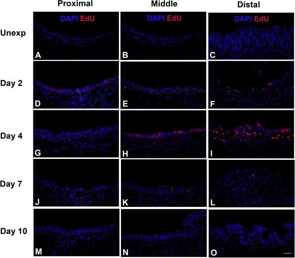
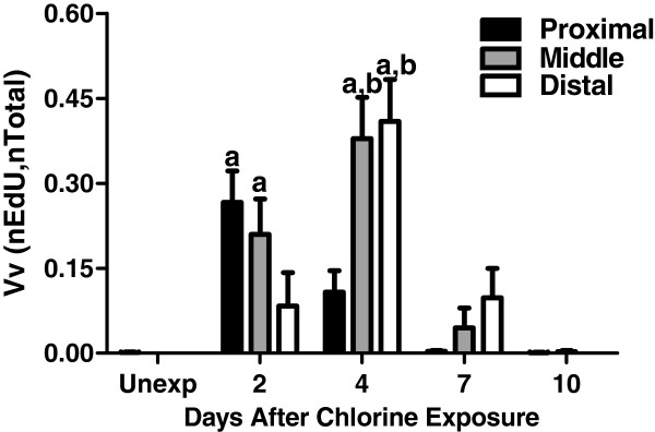
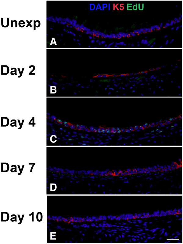
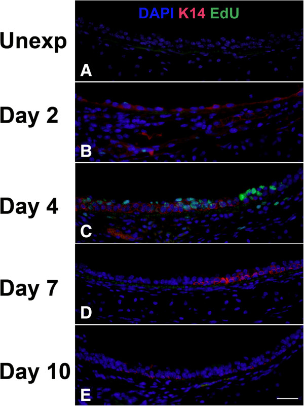
C57BL/6 mice were exposed to chlorine and injected with 5-ethynyl-2'-deoxyuridine (EdU) to label proliferating cells prior to sacrifice and collection of tracheas on days 2, 4, 7, and 10 after exposure. Airway repair and restoration of a differentiated epithelium were examined by co-localization of EdU labeling with markers for the three major tracheal epithelial cell types [keratin 5 (K5) and keratin 14 (K14) for basal cells, Clara cell secretory protein (CCSP) for Clara cells, and acetylated tubulin (AcTub) for ciliated cells]. Morphometric analysis was used to measure proliferation and restoration of a pseudostratified epithelium.
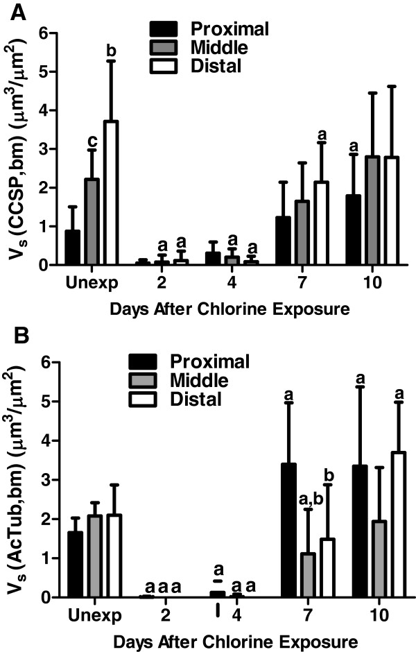
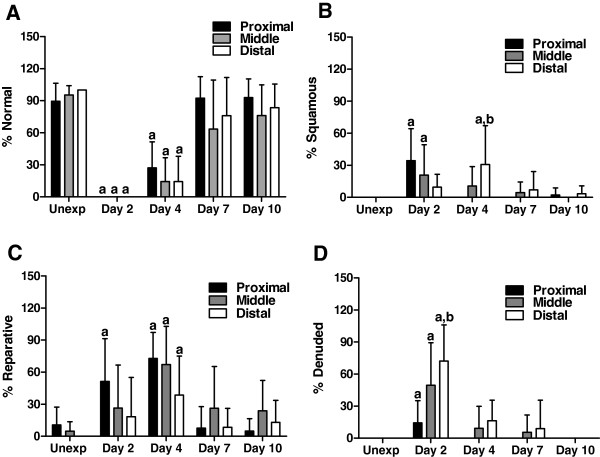
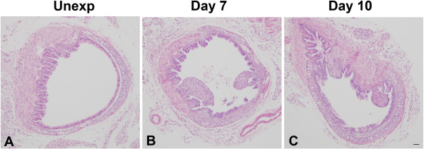
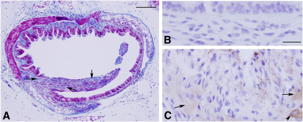
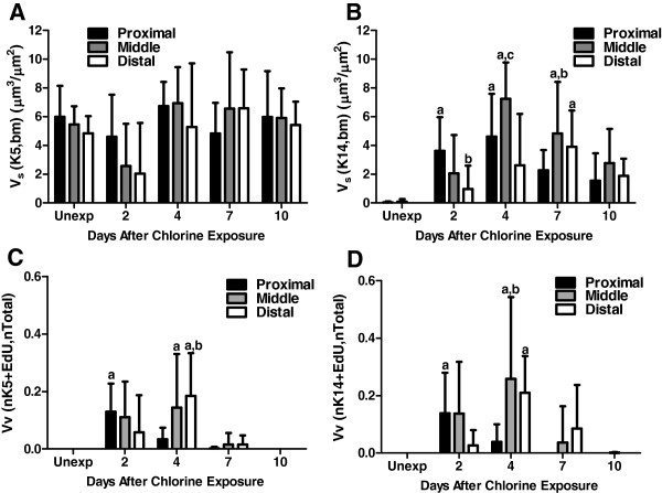
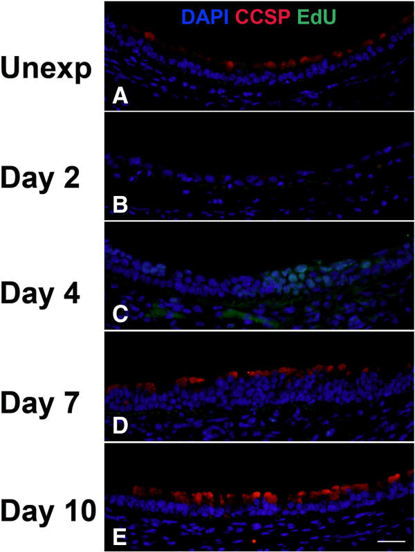
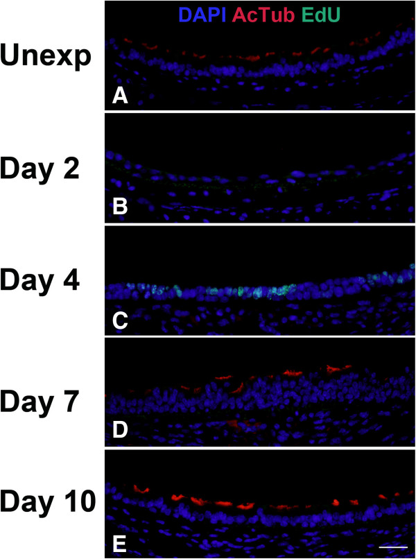
Epithelial repair was fastest and most extensive in proximal trachea compared with middle and distal trachea. In unexposed mice, cell proliferation was minimal, all basal cells expressed K5, and K14-expressing basal cells were absent from most sections. Chlorine exposure resulted in the sloughing of Clara and ciliated cells from the tracheal epithelium. Two to four days after chlorine exposure, cell proliferation occurred in K5- and K14-expressing basal cells, and the number of K14 cells was dramatically increased. In the period of peak cell proliferation, few if any ciliated or Clara cells were detected in repairing trachea. Expression of ciliated and Clara cell markers was detected at later times (days 7-10), but cell proliferation was not detected in areas in which these differentiated markers were re-expressed. Fibrotic lesions were observed at days 7-10 primarily in distal trachea.
The data are consistent with a model where surviving basal cells function as progenitor cells to repopulate the tracheal epithelium after chlorine injury. In areas with few remaining basal cells, repair is inefficient, leading to airway fibrosis. These studies establish a model for understanding regenerative processes in the respiratory epithelium useful for testing therapies for airway injury.
氯气是一种广泛使用的有毒化合物,被认为是一种化学威胁剂。氯气吸入会损伤气道上皮细胞,导致肺部异常。有效修复受损的上皮细胞对于恢复正常的肺结构和功能是必要的。本研究的目的是描述急性氯气损伤后气管上皮的修复过程。
将 C57BL/6 小鼠暴露于氯气中,并在暴露后第 2、4、7 和 10 天处死小鼠,收集气管,并用 5-乙炔基-2'-脱氧尿苷(EdU)标记增殖细胞。通过 EdU 标记与三种主要气管上皮细胞类型(基底细胞的角蛋白 5(K5)和角蛋白 14(K14)、Clara 细胞分泌蛋白(CCSP)和乙酰化微管蛋白(AcTub)的共定位,检测气道修复和分化上皮的恢复情况。形态计量分析用于测量增殖和假复层上皮的恢复。
与中、远段气管相比,近端气管的上皮修复最快、最广泛。在未暴露的小鼠中,细胞增殖很少,所有基底细胞均表达 K5,并且 K14 表达的基底细胞在大多数切片中均不存在。氯气暴露导致 Clara 和纤毛细胞从气管上皮脱落。氯气暴露后 2-4 天,K5 和 K14 表达的基底细胞发生细胞增殖,K14 细胞数量显著增加。在细胞增殖高峰期,很少有纤毛细胞或 Clara 细胞在修复的气管中被检测到。在稍后的时间(第 7-10 天)检测到纤毛细胞和 Clara 细胞标记物的表达,但在这些分化标记物重新表达的区域未检测到细胞增殖。第 7-10 天主要在远段气管观察到纤维性病变。
这些数据与一种模型一致,即存活的基底细胞作为祖细胞,在氯气损伤后重新填充气管上皮。在剩余基底细胞较少的区域,修复效率较低,导致气道纤维化。这些研究建立了一种理解呼吸上皮再生过程的模型,对于测试气道损伤的治疗方法很有用。