Department of Biomedical and NeuroMotor Sciences (DiBiNeM), University of Bologna, Bologna, Italy.
PLoS One. 2012;7(11):e50230. doi: 10.1371/journal.pone.0050230. Epub 2012 Nov 27.
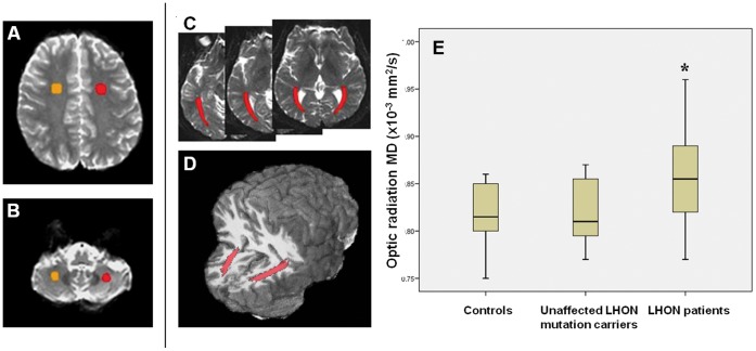
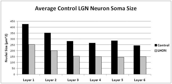
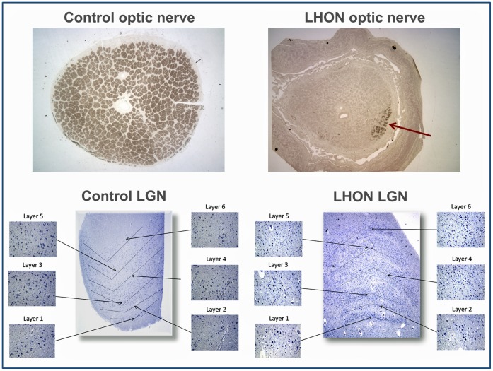
Leber's hereditary optic neuropathy (LHON) is characterized by retinal ganglion cell (RGC) degeneration with the preferential involvement of those forming the papillomacular bundle. The optic nerve is considered the main pathological target for LHON. Our aim was to investigate the possible involvement of the post-geniculate visual pathway in LHON patients. We used diffusion-weighted imaging for in vivo evaluation. Mean diffusivity maps from 22 LHON visually impaired, 11 unaffected LHON mutation carriers and 22 healthy subjects were generated and compared at level of optic radiation (OR). Prefrontal and cerebellar white matter were also analyzed as internal controls. Furthermore, we studied the optic nerve and the lateral geniculate nucleus (LGN) in post-mortem specimens obtained from a severe case of LHON compared to an age-matched control. Mean diffusivity values of affected patients were higher than unaffected mutation carriers (P<0.05) and healthy subjects (P<0.01) in OR and not in the other brain regions. Increased OR diffusivity was associated with both disease duration (B = 0.002; P<0.05) and lack of recovery of visual acuity (B = 0.060; P<0.01). Post-mortem investigation detected atrophy (41.9% decrease of neuron soma size in the magnocellular layers and 44.7% decrease in the parvocellular layers) and, to a lesser extent, degeneration (28.5% decrease of neuron density in the magnocellular layers and 28.7% decrease in the parvocellular layers) in the LHON LGN associated with extremely severe axonal loss (99%) in the optic nerve. The post-geniculate involvement in LHON patients is a downstream post-synaptic secondary phenomenon, reflecting de-afferentation rather than a primary neurodegeneration due to mitochondrial dysfunction of LGN neurons.
Leber 遗传性视神经病变 (LHON) 的特征是视网膜神经节细胞 (RGC) 变性,优先累及形成视乳头黄斑束的细胞。视神经被认为是 LHON 的主要病理靶标。我们的目的是研究 LHON 患者的视路后段是否可能受累。我们使用扩散加权成像进行体内评估。生成了 22 名 LHON 视力障碍患者、11 名未受影响的 LHON 突变携带者和 22 名健康对照者的视辐射 (OR) 平均弥散度图,并进行了比较。还分析了前额叶和小脑白质作为内部对照。此外,我们研究了从一例严重 LHON 病例获得的视神经和外侧膝状体 (LGN) 与年龄匹配的对照进行了比较。受影响患者的 OR 平均弥散度值高于未受影响的突变携带者 (P<0.05) 和健康对照组 (P<0.01),而其他脑区则没有。OR 弥散度的增加与疾病持续时间 (B=0.002;P<0.05) 和视力恢复不良 (B=0.060;P<0.01) 有关。尸检研究发现 LHON 的 LGN 存在萎缩(大细胞层神经元胞体大小减少 41.9%,小细胞层减少 44.7%)和退行性变(大细胞层神经元密度减少 28.5%,小细胞层减少 28.7%),与视神经中极为严重的轴突丢失(99%)相关。LHON 患者的视路后段受累是一种下游的突触后继发性现象,反映了去传入而不是由于 LGN 神经元的线粒体功能障碍导致的原发性神经退行性变。