Department of Pharmacology and Toxicology, Virginia Commonwealth University, Medical College of Virginia Campus, Richmond, Virginia 23298, USA.
Biol Psychiatry. 2013 Mar 1;73(5):443-53. doi: 10.1016/j.biopsych.2012.09.026. Epub 2012 Dec 4.
Human immunodeficiency virus (HIV) associated neurocognitive disorders (HAND), including memory dysfunction, continue to be a major clinical manifestation of HIV type-1 infection. Viral proteins released by infected glia are thought to be the principal triggers of inflammation and bystander neuronal injury and death, thereby driving key symptomatology of HAND.
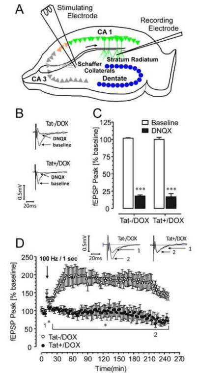
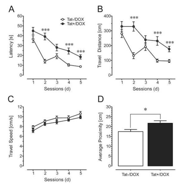
We used a glial fibrillary acidic protein-driven, doxycycline-inducible HIV type-1 transactivator of transcription (Tat) transgenic mouse model and examined structure-function relationships in hippocampal pyramidal cornu ammonis 1 (CA1) neurons using morphologic, electrophysiological (long-term potentiation [LTP]), and behavioral (Morris water maze, fear-conditioning) approaches.
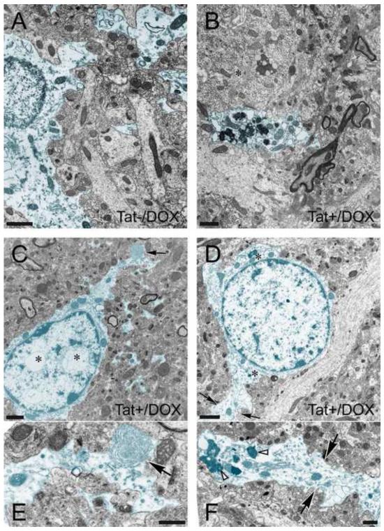
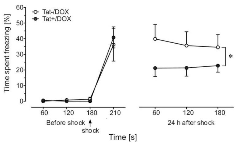
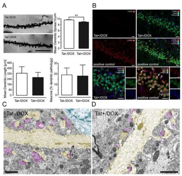
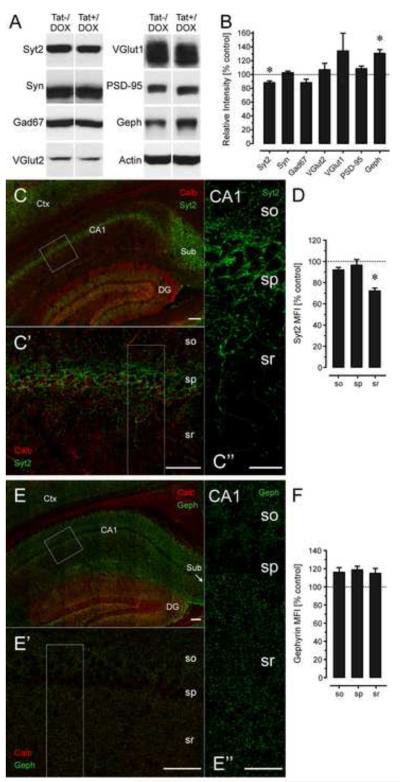
Tat induction caused a variety of different inclusions in astrocytes characteristic of lysosomes, autophagic vacuoles, and lamellar bodies, which were typically present within distal cytoplasmic processes. In pyramidal CA1 neurons, Tat induction reduced the number of apical dendritic spines, while disrupting the distribution of synaptic proteins (synaptotagmin 2 and gephyrin) associated with inhibitory transmission but with minimal dendritic pathology and no evidence of pyramidal neuron death. Electrophysiological assessment of excitatory postsynaptic field potential at Schaffer collateral/commissural fiber-CA1 synapses showed near total suppression of LTP in mice expressing Tat. The loss in LTP coincided with disruptions in learning and memory.
Tat expression in the brain results in profound functional changes in synaptic physiology and in behavior that are accompanied by only modest structural changes and minimal pathology. Tat likely contributes to HAND by causing molecular changes that disrupt synaptic organization, with inhibitory presynaptic terminals containing synaptotagmin 2 appearing especially vulnerable.
人类免疫缺陷病毒(HIV)相关的神经认知障碍(HAND),包括记忆功能障碍,仍然是 HIV 型 1 感染的主要临床表现。受感染的神经胶质细胞释放的病毒蛋白被认为是引发炎症和旁观者神经元损伤和死亡的主要触发因素,从而推动 HAND 的关键症状。
我们使用胶质纤维酸性蛋白驱动的、强力霉素诱导的 HIV 型 1 转录激活剂(Tat)转基因小鼠模型,并用形态学、电生理学(长时程增强[LTP])和行为学(Morris 水迷宫、恐惧条件反射)方法研究海马角状突 1(CA1)神经元的结构-功能关系。
Tat 诱导导致星形胶质细胞中出现各种不同的包含物,具有溶酶体、自噬空泡和板层小体的特征,这些包含物通常存在于远端细胞质突起内。在 CA1 锥体神经元中,Tat 诱导减少了树突棘的数量,同时破坏了与抑制性传递相关的突触蛋白(突触结合蛋白 2 和 Gephyrin)的分布,但树突病理学最小,没有锥体神经元死亡的证据。在表达 Tat 的小鼠中,兴奋性突触后场电位的电生理评估显示 Schaffer 侧支/联合纤维-CA1 突触的 LTP 几乎完全抑制。LTP 的丧失与学习和记忆的障碍相吻合。
大脑中的 Tat 表达导致突触生理学和行为方面的深刻功能变化,只有适度的结构变化和最小的病理学。Tat 可能通过引起破坏突触组织的分子变化导致 HAND,其中含有突触结合蛋白 2 的抑制性突触前末端显得特别脆弱。