Kalokairinou Kiriaki, Konstantinidis Charalampos, Domazou Marilena, Kalogeropoulos Theodoros, Kosmidis Prodromos, Gekas Aristomenis
Department of Radiology, National Rehabilitation Center, Ilion, Athens, Greece.
J Clin Imaging Sci. 2012;2:63. doi: 10.4103/2156-7514.103053. Epub 2012 Oct 31.
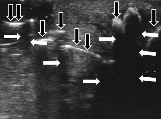
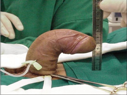
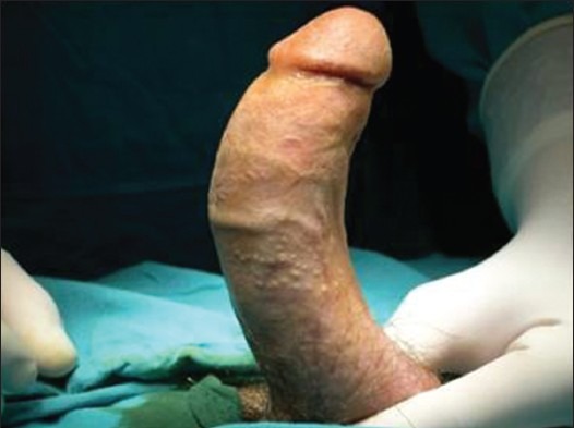
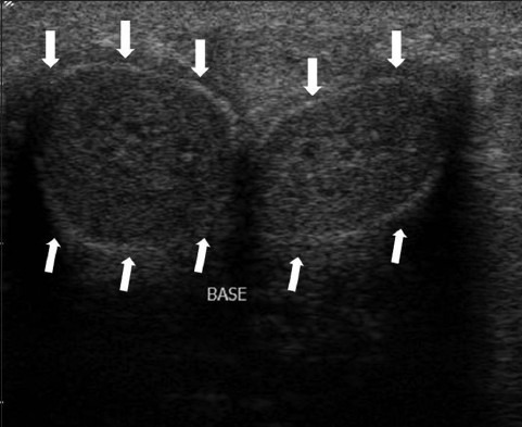
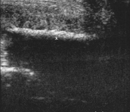
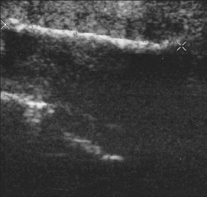
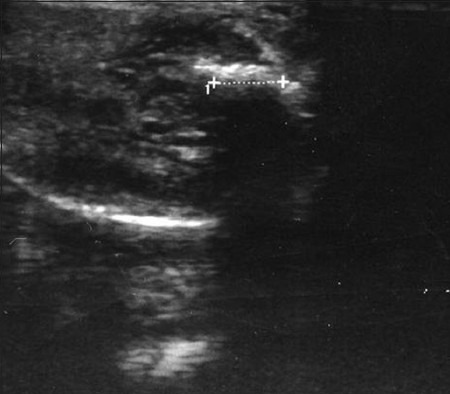
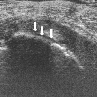
The aim of this study is to assess the role of ultrasound (US) in Peyronie's Disease (PD). PD is a psychologically and physically devastating disorder that manifests in middle-aged men. Fibrous inelastic plaques in the tunica albuginea, result in palpable penile scar in the flaccid condition and cause painful erections and penile deformity, including penile curvature, hinging, narrowing, and shortening of penis. Penile deformity is the most common (52%) first symptom of PD and is present in 94% of affected men. US is the primary imaging modality of choice due to its easy availability, low risk, and ability to image and quantify both calcified and soft tissue elements of PD. US provides identification of smaller and non-palpable lesions and shows the extent of fibrosis. Detection of calcifications within the plaque suggests stabilization of the disease and provides information useful to select patients for appropriate treatment.
本研究的目的是评估超声(US)在佩罗尼氏病(PD)中的作用。PD是一种对心理和身体都具有破坏性的疾病,多见于中年男性。白膜中的纤维性无弹性斑块,在阴茎疲软状态下可触及阴茎瘢痕,并导致疼痛性勃起和阴茎畸形,包括阴茎弯曲、铰链样改变、变窄和阴茎缩短。阴茎畸形是PD最常见的首发症状(52%),94%的患病男性存在该症状。由于超声易于获得、风险低,且能够对PD的钙化和软组织成分进行成像和量化,因此它是首选的主要成像方式。超声可识别较小的、无法触及的病变,并显示纤维化程度。斑块内钙化的检测提示疾病已稳定,并为选择合适治疗的患者提供有用信息。