• 文献检索
  • 文档翻译
  • 深度研究
  • 学术资讯
  • Suppr Zotero 插件Zotero 插件
  • 邀请有礼
  • 套餐&价格
  • 历史记录
应用&插件
Suppr Zotero 插件Zotero 插件浏览器插件Mac 客户端Windows 客户端微信小程序
定价
高级版会员购买积分包购买API积分包
服务
文献检索文档翻译深度研究API 文档MCP 服务
关于我们
关于 Suppr公司介绍联系我们用户协议隐私条款
关注我们

Suppr 超能文献

核心技术专利:CN118964589B侵权必究
粤ICP备2023148730 号-1Suppr @ 2026

文献检索

告别复杂PubMed语法,用中文像聊天一样搜索,搜遍4000万医学文献。AI智能推荐,让科研检索更轻松。

立即免费搜索

文件翻译

保留排版,准确专业,支持PDF/Word/PPT等文件格式,支持 12+语言互译。

免费翻译文档

深度研究

AI帮你快速写综述,25分钟生成高质量综述,智能提取关键信息,辅助科研写作。

立即免费体验

通过丝裂原活化蛋白激酶激酶/细胞外信号调节激酶通路调节实验性蛛网膜下腔出血中促炎介质的增强性脑血管表达。

Regulation of enhanced cerebrovascular expression of proinflammatory mediators in experimental subarachnoid hemorrhage via the mitogen-activated protein kinase kinase/extracellular signal-regulated kinase pathway.

机构信息

Department of Clinical Sciences, Division of Experimental Vascular Research, Lund University, Lund, Sweden.

出版信息

J Neuroinflammation. 2012 Dec 21;9:274. doi: 10.1186/1742-2094-9-274.

DOI:10.1186/1742-2094-9-274
PMID:23259581
原文链接:https://pmc.ncbi.nlm.nih.gov/articles/PMC3573995/
Abstract

BACKGROUND

Subarachnoid hemorrhage (SAH) is associated with high morbidity and mortality. It is suggested that the associated inflammation is mediated through activation of the mitogen-activated protein kinase (MAPK) pathway which plays a crucial role in the pathogenesis of delayed cerebral ischemia after SAH. The aim of this study was first to investigate the timecourse of altered expression of proinflammatory cytokines and matrix metalloproteinase in the cerebral arteries walls following SAH. Secondly, we investigated whether administration of a specific mitogen-activated protein kinase kinase (MEK)1/2 inhibitor, U0126, given at 6 h after SAH prevents activation of the MEK/extracellular signal-regulated kinase 1/2 pathway and the upregulation of cerebrovascular inflammatory mediators and improves neurological function.

METHODS

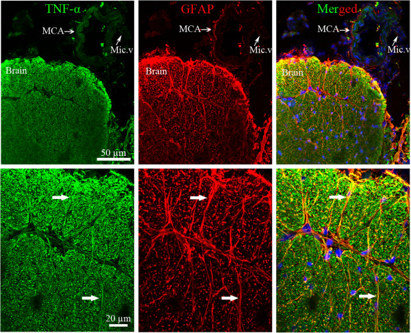
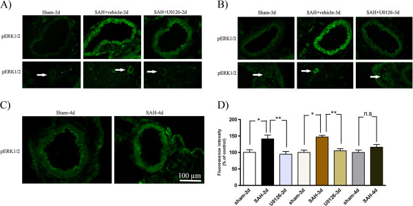
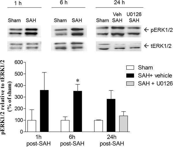
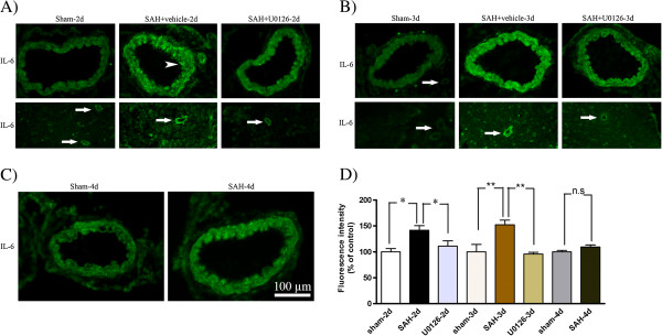
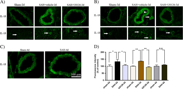
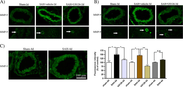
SAH was induced in rats by injection of 250 μl of autologous blood into basal cisterns. U0126 was given intracisternally using two treatment regimens: (A) treatments at 6, 12, 24 and 36 h after SAH and experiments terminated at 48 h after SAH, or (B) treatments at 6, 12, and 24 h after SAH and terminated at 72 h after SAH. Cerebral arteries were harvested and interleukin (IL)-6, IL-1β, tumor necrosis factor α (TNF)α, matrix metalloproteinase (MMP)-9 and phosphorylated ERK1/2 (pERK1/2) levels investigated by immunohistochemistry. Early activation of pERK1/2 was measured by western blot. Functional neurological outcome after SAH was also analyzed.

RESULTS

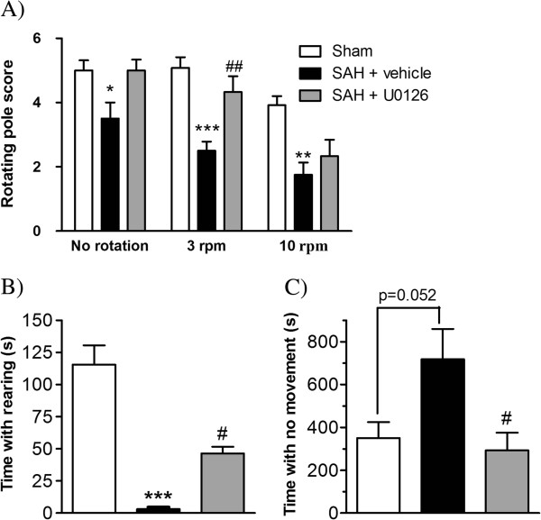
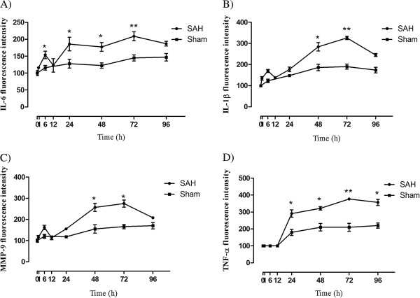
Expression levels of IL-1β, IL-6, MMP-9 and pERK1/2 proteins were elevated over time with an early increase at around 6 h and a late peak at 48 to 72 h post-SAH in cerebral arteries. Enhanced expression of TNFα in cerebral arteries started at 24 h and increased until 96 h. In addition, SAH induced sensorimotor and spontaneous behavior deficits in the animals. Treatment with U0126 starting at 6 h after SAH prevented activation of MEK-ERK1/2 signaling. Further, U0126 significantly decreased the upregulation of inflammation proteins at 48 and 72 h following SAH and improved neurological function. We found no differences between treatment regimens A and B.

CONCLUSIONS

These results show that SAH induces early activation of the MEK-ERK1/2 pathway in cerebral artery walls, which is associated with upregulation of proinflammatory cytokines and MMP-9. Inhibition of the MEK-ERK1/2 pathway by U0126 starting at 6 h post-SAH prevented upregulation of cytokines and MMP-9 in cerebral vessels, and improved neurological outcome.

摘要

背景

蛛网膜下腔出血(SAH)与高发病率和死亡率相关。据认为,相关炎症是通过丝裂原活化蛋白激酶(MAPK)途径的激活介导的,MAPK 途径在蛛网膜下腔出血后迟发性脑缺血的发病机制中起关键作用。本研究的目的首先是研究蛛网膜下腔出血后动脉壁中促炎细胞因子和基质金属蛋白酶表达的时间变化。其次,我们研究了在蛛网膜下腔出血后 6 小时给予特定的丝裂原活化蛋白激酶激酶(MEK)1/2 抑制剂 U0126 是否可以防止 MEK/细胞外信号调节激酶 1/2 途径的激活以及脑血管炎症介质的上调,并改善神经功能。

方法

通过将 250μl 自体血液注入基底池来诱导大鼠蛛网膜下腔出血。通过两种治疗方案进行 U0126 鞘内给药:(A)蛛网膜下腔出血后 6、12、24 和 36 小时给予治疗,实验在蛛网膜下腔出血后 48 小时终止,或(B)蛛网膜下腔出血后 6、12 和 24 小时给予治疗,在蛛网膜下腔出血后 72 小时终止。通过免疫组织化学法研究白细胞介素(IL)-6、IL-1β、肿瘤坏死因子α(TNF)α、基质金属蛋白酶(MMP)-9 和磷酸化 ERK1/2(pERK1/2)水平。通过 Western blot 测定 pERK1/2 的早期激活。还分析了蛛网膜下腔出血后的早期神经功能。

结果

IL-1β、IL-6、MMP-9 和 pERK1/2 蛋白的表达水平随时间推移而升高,在蛛网膜下腔出血后约 6 小时出现早期增加,在 48 至 72 小时出现晚期高峰。在大脑动脉中,TNFα的增强表达在 24 小时开始并增加直至 96 小时。此外,蛛网膜下腔出血诱导动物出现感觉运动和自发性行为缺陷。在蛛网膜下腔出血后 6 小时开始用 U0126 治疗可防止 MEK-ERK1/2 信号的激活。进一步,U0126 显著降低了蛛网膜下腔出血后 48 小时和 72 小时炎症蛋白的上调,并改善了神经功能。我们在治疗方案 A 和 B 之间未发现差异。

结论

这些结果表明,蛛网膜下腔出血诱导大脑动脉壁中 MEK-ERK1/2 途径的早期激活,这与促炎细胞因子和 MMP-9 的上调有关。在蛛网膜下腔出血后 6 小时开始用 U0126 抑制 MEK-ERK1/2 途径可防止血管中细胞因子和 MMP-9 的上调,并改善神经功能。

https://cdn.ncbi.nlm.nih.gov/pmc/blobs/b567/3573995/9ec82db0d4a1/1742-2094-9-274-10.jpg
https://cdn.ncbi.nlm.nih.gov/pmc/blobs/b567/3573995/a49b2d2d36dc/1742-2094-9-274-1.jpg
https://cdn.ncbi.nlm.nih.gov/pmc/blobs/b567/3573995/7b804210d648/1742-2094-9-274-2.jpg
https://cdn.ncbi.nlm.nih.gov/pmc/blobs/b567/3573995/08c3fad44321/1742-2094-9-274-3.jpg
https://cdn.ncbi.nlm.nih.gov/pmc/blobs/b567/3573995/15a8995815bb/1742-2094-9-274-4.jpg
https://cdn.ncbi.nlm.nih.gov/pmc/blobs/b567/3573995/2daaa217ab50/1742-2094-9-274-5.jpg
https://cdn.ncbi.nlm.nih.gov/pmc/blobs/b567/3573995/5ebeef26054e/1742-2094-9-274-6.jpg
https://cdn.ncbi.nlm.nih.gov/pmc/blobs/b567/3573995/4a7f820f85c5/1742-2094-9-274-7.jpg
https://cdn.ncbi.nlm.nih.gov/pmc/blobs/b567/3573995/316c313a3945/1742-2094-9-274-8.jpg
https://cdn.ncbi.nlm.nih.gov/pmc/blobs/b567/3573995/28ebe92168c3/1742-2094-9-274-9.jpg
https://cdn.ncbi.nlm.nih.gov/pmc/blobs/b567/3573995/9ec82db0d4a1/1742-2094-9-274-10.jpg

相似文献

1
Regulation of enhanced cerebrovascular expression of proinflammatory mediators in experimental subarachnoid hemorrhage via the mitogen-activated protein kinase kinase/extracellular signal-regulated kinase pathway.通过丝裂原活化蛋白激酶激酶/细胞外信号调节激酶通路调节实验性蛛网膜下腔出血中促炎介质的增强性脑血管表达。
J Neuroinflammation. 2012 Dec 21;9:274. doi: 10.1186/1742-2094-9-274.
2
Improvement in neurological outcome and abolition of cerebrovascular endothelin B and 5-hydroxytryptamine 1B receptor upregulation through mitogen-activated protein kinase kinase 1/2 inhibition after subarachnoid hemorrhage in rats.大鼠蛛网膜下腔出血后通过丝裂原活化蛋白激酶激酶 1/2 抑制改善神经功能预后和消除脑血管内皮素 B 和 5-羟色胺 1B 受体上调。
J Neurosurg. 2011 Apr;114(4):1143-53. doi: 10.3171/2010.6.JNS1018. Epub 2010 Jul 2.
3
Blockade of the MEK/ERK pathway with a raf inhibitor prevents activation of pro-inflammatory mediators in cerebral arteries and reduction in cerebral blood flow after subarachnoid hemorrhage in a rat model.RAF 抑制剂阻断 MEK/ERK 通路可预防蛛网膜下腔出血大鼠模型脑血管中促炎介质的激活和脑血流减少。
J Cereb Blood Flow Metab. 2011 Jan;31(1):144-54. doi: 10.1038/jcbfm.2010.62. Epub 2010 Apr 28.
4
Proteomic Expression Changes in Large Cerebral Arteries After Experimental Subarachnoid Hemorrhage in Rat Are Regulated by the MEK-ERK1/2 Pathway.大鼠实验性蛛网膜下腔出血后大脑大动脉中的蛋白质组表达变化受MEK-ERK1/2信号通路调控。
J Mol Neurosci. 2017 Aug;62(3-4):380-394. doi: 10.1007/s12031-017-0944-7. Epub 2017 Jul 24.
5
Early events triggering delayed vasoconstrictor receptor upregulation and cerebral ischemia after subarachnoid hemorrhage.蛛网膜下腔出血后延迟性血管收缩受体上调和脑缺血的早期触发事件。
BMC Neurosci. 2013 Mar 15;14:34. doi: 10.1186/1471-2202-14-34.
6
Cerebral ischemia induces microvascular pro-inflammatory cytokine expression via the MEK/ERK pathway.脑缺血通过 MEK/ERK 通路诱导微血管促炎细胞因子表达。
J Neuroinflammation. 2010 Feb 26;7:14. doi: 10.1186/1742-2094-7-14.
7
ERK1/2 inhibition attenuates cerebral blood flow reduction and abolishes ET(B) and 5-HT(1B) receptor upregulation after subarachnoid hemorrhage in rat.抑制细胞外信号调节激酶1/2可减轻大鼠蛛网膜下腔出血后脑血流量的减少,并消除内皮素B型受体(ET(B))和5-羟色胺1B型受体(5-HT(1B))的上调。
J Cereb Blood Flow Metab. 2006 Jun;26(6):846-56. doi: 10.1038/sj.jcbfm.9600236.
8
Subtype activation and interaction of protein kinase C and mitogen-activated protein kinase controlling receptor expression in cerebral arteries and microvessels after subarachnoid hemorrhage.蛛网膜下腔出血后蛋白激酶C和丝裂原活化蛋白激酶的亚型激活及相互作用对脑动脉和微血管中受体表达的调控
Stroke. 2008 Jan;39(1):185-90. doi: 10.1161/STROKEAHA.107.487827. Epub 2007 Nov 21.
9
Enhanced cerebrovascular expression of matrix metalloproteinase-9 and tissue inhibitor of metalloproteinase-1 via the MEK/ERK pathway during cerebral ischemia in the rat.大鼠脑缺血期间通过MEK/ERK途径增强基质金属蛋白酶-9和金属蛋白酶组织抑制剂-1的脑血管表达。
BMC Neurosci. 2009 Jun 4;10:56. doi: 10.1186/1471-2202-10-56.
10
CaMKII inhibition with KN93 attenuates endothelin and serotonin receptor-mediated vasoconstriction and prevents subarachnoid hemorrhage-induced deficits in sensorimotor function.用KN93抑制钙/钙调蛋白依赖性蛋白激酶II可减轻内皮素和5-羟色胺受体介导的血管收缩,并预防蛛网膜下腔出血引起的感觉运动功能缺陷。
J Neuroinflammation. 2014 Dec 10;11:207. doi: 10.1186/s12974-014-0207-2.

引用本文的文献

1
CDDO, an Anti-Inflammatory and Antioxidant Compound, Attenuates Vasospasm and Neuronal Cell Apoptosis in Rats Subjected to Experimental Subarachnoid Hemorrhage.CDDO,一种抗炎和抗氧化化合物,可减轻实验性蛛网膜下腔出血大鼠的血管痉挛和神经元细胞凋亡。
Curr Issues Mol Biol. 2024 May 13;46(5):4688-4700. doi: 10.3390/cimb46050283.
2
All Three Supersystems-Nervous, Vascular, and Immune-Contribute to the Cortical Infarcts After Subarachnoid Hemorrhage.神经系统、血管系统和免疫系统这三大超级系统均与蛛网膜下腔出血后的皮质梗死有关。
Transl Stroke Res. 2025 Feb;16(1):96-118. doi: 10.1007/s12975-024-01242-z. Epub 2024 Apr 30.
3
Switching Off Vascular MAPK Signaling: A Novel Strategy to Prevent Delayed Cerebral Ischemia Following Subarachnoid Hemorrhage.

本文引用的文献

1
Potential contribution of hypoxia-inducible factor-1α, aquaporin-4, and matrix metalloproteinase-9 to blood-brain barrier disruption and brain edema after experimental subarachnoid hemorrhage.实验性蛛网膜下腔出血后血脑屏障破坏和脑水肿中缺氧诱导因子-1α、水通道蛋白-4 和基质金属蛋白酶-9 的潜在作用。
J Mol Neurosci. 2012 Sep;48(1):273-80. doi: 10.1007/s12031-012-9769-6. Epub 2012 Apr 22.
2
Matrix metalloproteinase-9 concentration in the cerebral extracellular fluid of patients during the acute phase of aneurysmal subarachnoid hemorrhage.动脉瘤性蛛网膜下腔出血急性期患者脑细胞外液中基质金属蛋白酶-9的浓度
Neurol Res. 2012 Jun;34(5):455-61. doi: 10.1179/1743132812Y.0000000018. Epub 2012 Mar 20.
3
关闭血管丝裂原活化蛋白激酶信号传导:预防蛛网膜下腔出血后迟发性脑缺血的新策略。
Transl Stroke Res. 2025 Jun;16(3):952-961. doi: 10.1007/s12975-024-01234-z. Epub 2024 Feb 9.
4
Ravoxertinib Improves Long-Term Neurologic Deficits after Experimental Subarachnoid Hemorrhage through Early Inhibition of Erk1/2.瑞伏昔替尼通过早期抑制Erk1/2改善实验性蛛网膜下腔出血后的长期神经功能缺损。
ACS Omega. 2023 May 23;8(22):19692-19704. doi: 10.1021/acsomega.3c01296. eCollection 2023 Jun 6.
5
Immunological Profile of Vasospasm after Subarachnoid Hemorrhage.蛛网膜下腔出血后血管痉挛的免疫特征。
Int J Mol Sci. 2023 May 16;24(10):8856. doi: 10.3390/ijms24108856.
6
Diffuse noxious inhibitory controls in chronic joint inflammatory Pain: Study of the descending serotonergic modulation mediated through 5HT3 receptors.慢性关节炎性疼痛中的弥漫性伤害性抑制控制:通过5-羟色胺3(5HT3)受体介导的下行5-羟色胺能调制的研究
Neurobiol Pain. 2023 Mar 1;13:100123. doi: 10.1016/j.ynpai.2023.100123. eCollection 2023 Jan-Jul.
7
Overexpression of mitogen-activated protein kinase phosphatase-1 in endothelial cells reduces blood-brain barrier injury in a mouse model of ischemic stroke.丝裂原活化蛋白激酶磷酸酶-1在内皮细胞中的过表达可减轻缺血性脑卒中小鼠模型的血脑屏障损伤。
Neural Regen Res. 2023 Aug;18(8):1743-1749. doi: 10.4103/1673-5374.363836.
8
Inflammation and immune cell abnormalities in intracranial aneurysm subarachnoid hemorrhage (SAH): Relevant signaling pathways and therapeutic strategies.颅内动脉瘤性蛛网膜下腔出血(SAH)中的炎症和免疫细胞异常:相关信号通路和治疗策略。
Front Immunol. 2022 Nov 23;13:1027756. doi: 10.3389/fimmu.2022.1027756. eCollection 2022.
9
Specific signature biomarkers highlight the potential mechanisms of circulating neutrophils in aneurysmal subarachnoid hemorrhage.特定的标志性生物标志物揭示了循环中性粒细胞在动脉瘤性蛛网膜下腔出血中的潜在机制。
Front Pharmacol. 2022 Nov 10;13:1022564. doi: 10.3389/fphar.2022.1022564. eCollection 2022.
10
Therapeutic Effect of Mitochondrial Division Inhibitor-1 (Mdivi-1) on Hyperglycemia-Exacerbated Early and Delayed Brain Injuries after Experimental Subarachnoid Hemorrhage.线粒体分裂抑制剂-1(Mdivi-1)对实验性蛛网膜下腔出血后高血糖加重的早期和迟发性脑损伤的治疗作用。
Int J Mol Sci. 2022 Jun 22;23(13):6924. doi: 10.3390/ijms23136924.
Potential contribution of matrix metalloproteinase-9 (mmp-9) to cerebral vasospasm after experimental subarachnoid hemorrhage in rats.
基质金属蛋白酶-9(MMP-9)对大鼠实验性蛛网膜下腔出血后脑血管痉挛的潜在作用。
Ann Clin Lab Sci. 2012 Winter;42(1):14-20.
4
Cell death starts early after subarachnoid hemorrhage.蛛网膜下腔出血后细胞死亡很早就开始了。
Neurosci Lett. 2012 Mar 14;512(1):6-11. doi: 10.1016/j.neulet.2012.01.036. Epub 2012 Jan 24.
5
Elevated peripheral neutrophils and matrix metalloproteinase 9 as biomarkers of functional outcome following subarachnoid hemorrhage.蛛网膜下腔出血后功能结局的生物标志物:外周血中性粒细胞和基质金属蛋白酶 9 升高。
Transl Stroke Res. 2011 Dec;2(4):600-7. doi: 10.1007/s12975-011-0117-x. Epub 2011 Nov 10.
6
Contribution of matrix metalloproteinase-9 to cerebral edema and functional outcome following experimental subarachnoid hemorrhage.基质金属蛋白酶-9 对实验性蛛网膜下腔出血后脑水肿和功能结果的影响。
Cerebrovasc Dis. 2011;32(3):289-95. doi: 10.1159/000328248. Epub 2011 Sep 9.
7
Aneurysmal subarachnoid hemorrhage: an update on the medical complications and treatments strategies seen in these patients.颅内动脉瘤性蛛网膜下腔出血:这些患者的医疗并发症及治疗策略的最新进展。
Curr Opin Anaesthesiol. 2011 Oct;24(5):500-7. doi: 10.1097/ACO.0b013e32834ad45b.
8
Vascular plasticity in cerebrovascular disorders.脑血管疾病中的血管可塑性。
J Cereb Blood Flow Metab. 2011 Jul;31(7):1554-71. doi: 10.1038/jcbfm.2011.70. Epub 2011 May 11.
9
Monitoring of the inflammatory response after aneurysmal subarachnoid haemorrhage in the clinical setting: review of literature and report of preliminary clinical experience.临床环境中动脉瘤性蛛网膜下腔出血后炎症反应的监测:文献综述及初步临床经验报告
Acta Neurochir Suppl. 2011;110(Pt 1):191-6. doi: 10.1007/978-3-7091-0353-1_33.
10
Mechanisms of early brain injury after SAH: matrix metalloproteinase 9.蛛网膜下腔出血后早期脑损伤的机制:基质金属蛋白酶9
Acta Neurochir Suppl. 2011;110(Pt 1):63-5. doi: 10.1007/978-3-7091-0353-1_11.