Department of Anatomy and Neurobiology, University of California, Irvine School of Medicine, 1001 Health Sciences Rd, 306D Med Surg II, Irvine, CA 92697, USA.
Cardiovasc Toxicol. 2013 Jun;13(2):161-7. doi: 10.1007/s12012-012-9194-7.
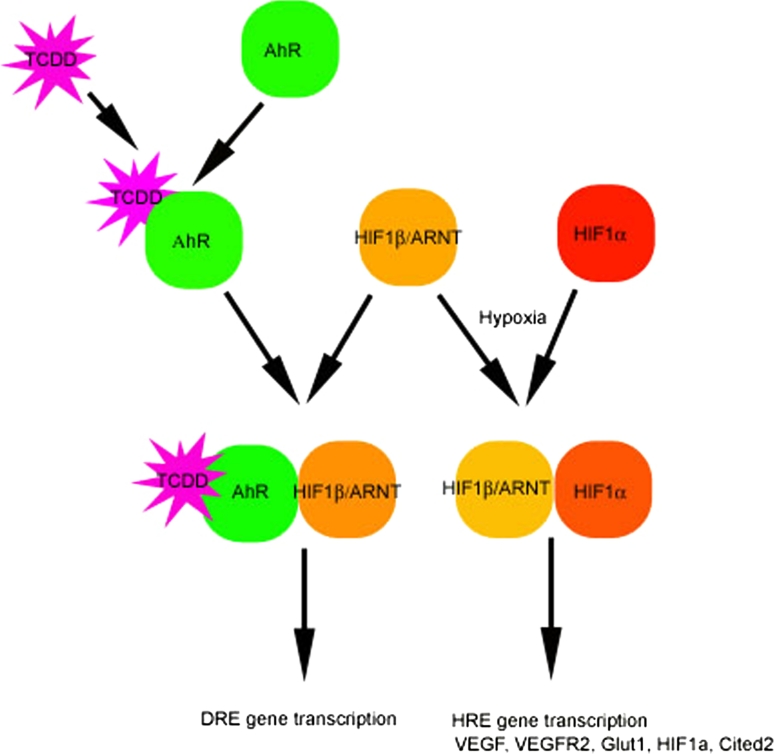
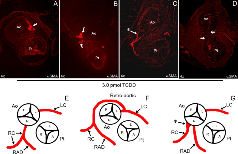
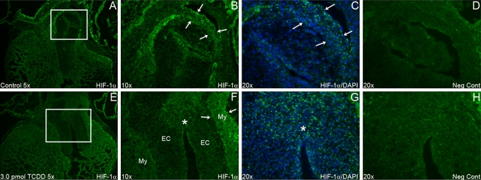
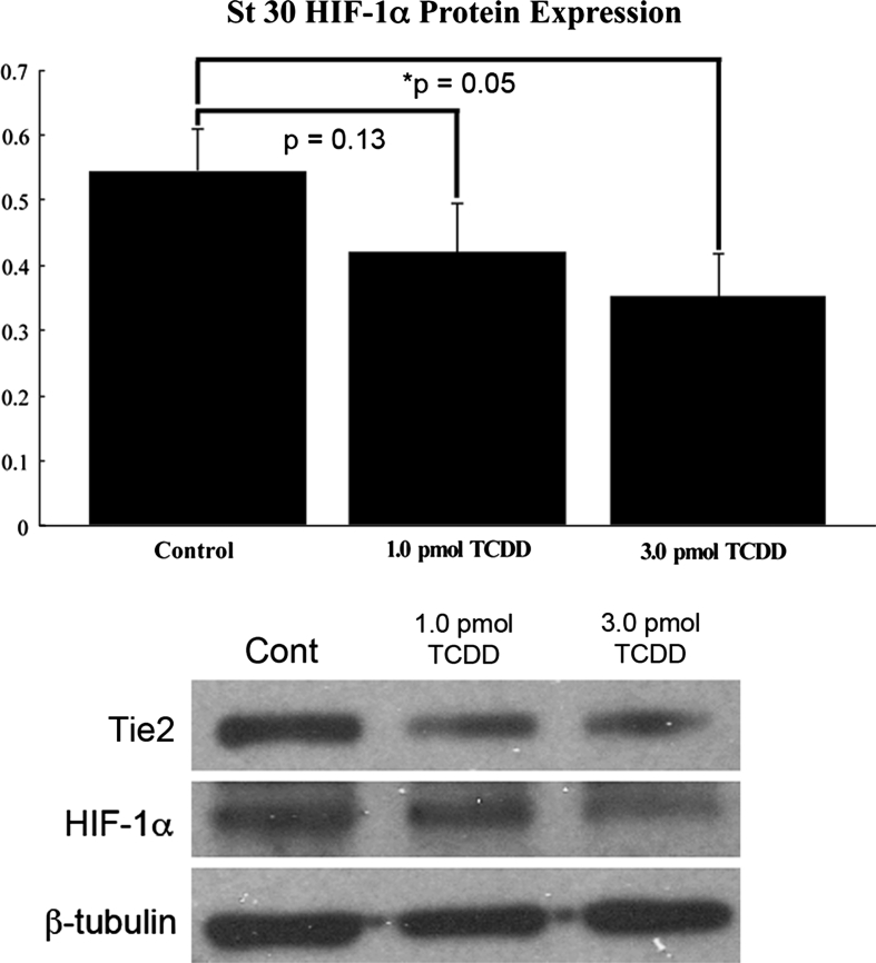
Differential tissue hypoxia drives normal cardiogenic events including coronary vessel development. This requirement renders cardiogenic processes potentially susceptible to teratogens that activate a transcriptional pathway that intersects with the hypoxia-inducible factor (HIF-1) pathway. The potent toxin 2,3,7,8-Tetrachlorodibenzo-p-dioxin (TCDD) is known to cause cardiovascular defects by way of reduced myocardial hypoxia, inhibition of angiogenic stimuli, and alterations in responsiveness of endothelial cells to those stimuli. Our working hypothesis is that HIF-1 levels and thus HIF-1 signaling in the developing myocardium will be reduced by TCDD treatment in vivo during a critical stage and in particularly sensitive sites during heart morphogenesis. This inadequate HIF-1 signaling will subsequently result in outflow tract (OFT) and coronary vasculature defects. Our current data using the chicken embryo model showed a marked decrease in the intensity of immunostaining for HIF-1α nuclear expression in the OFT myocardium of TCDD-treated embryos. This area at the base of the OFT is particularly hypoxic during normal development; where endothelial cells initially form a concentrated anastomosing network known as the peritruncal ring; and where the left and right coronary arteries eventually connect to the aortic lumen. Consistent with this finding, anomalies of the proximal coronaries were detected after TCDD treatment and HIF-1α protein levels decreased in a TCDD dose-dependent manner.
组织缺氧差异驱动正常的心发生事件,包括冠状血管发育。这种需求使心发生过程容易受到激活与缺氧诱导因子(HIF-1)途径相交的转录途径的致畸剂的影响。已知强力毒素 2,3,7,8-四氯二苯并对二恶英(TCDD)通过减少心肌缺氧、抑制血管生成刺激以及改变内皮细胞对这些刺激的反应性来引起心血管缺陷。我们的工作假设是,在心脏形态发生的关键阶段和特别敏感的部位,TCDD 在体内处理会降低发育中心肌中的 HIF-1 水平,从而降低 HIF-1 信号。这种不足的 HIF-1 信号随后会导致流出道(OFT)和冠状脉管系统缺陷。我们目前使用鸡胚模型的数据显示,TCDD 处理胚胎的 OFT 心肌中 HIF-1α核表达的免疫染色强度明显降低。OFT 底部的这个区域在正常发育过程中特别缺氧;内皮细胞最初形成一个密集的吻合网络,称为peri-truncal 环;左冠状动脉和右冠状动脉最终连接到主动脉腔。与这一发现一致,在 TCDD 处理后检测到近端冠状动脉的异常,并且 HIF-1α 蛋白水平以 TCDD 剂量依赖性方式降低。