Division of Gastroenterology, Department of Medicine, Taipei Veterans General Hospital and National Yang-Ming University, #201 Shih-Pai Road, Section 2, Taipei, Taiwan, ROC.
BMC Gastroenterol. 2012 Dec 29;12:182. doi: 10.1186/1471-230X-12-182.
The best sites for biopsy-based tests to evaluate H. pylori infection in gastritis with atrophy are not well known. This study aimed to evaluate the site and sensitivity of biopsy-based tests in terms of degree of gastritis with atrophy.
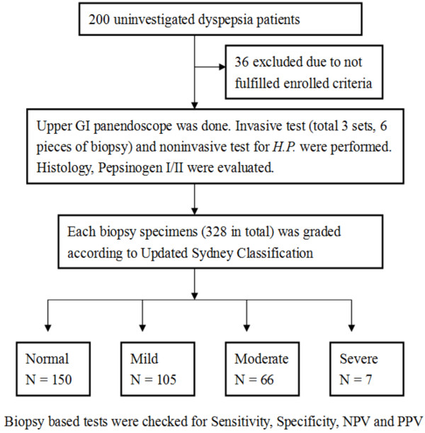
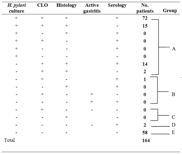
One hundred and sixty-four (164) uninvestigated dyspepsia patients were enrolled. Biopsy-based tests (i.e., culture, histology Giemsa stain and rapid urease test) and non-invasive tests (anti-H. pylori IgG) were performed. The gold standard of H. pylori infection was defined according to previous criteria. The sensitivity, specificity, positive predictive rate and negative predictive rate of biopsy-based tests at the gastric antrum and body were calculated in terms of degree of gastritis with atrophy.
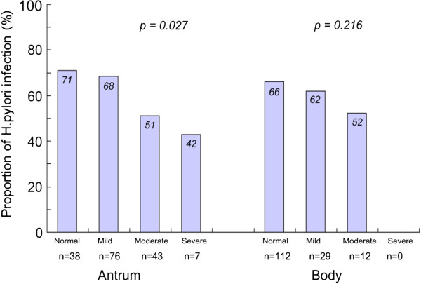
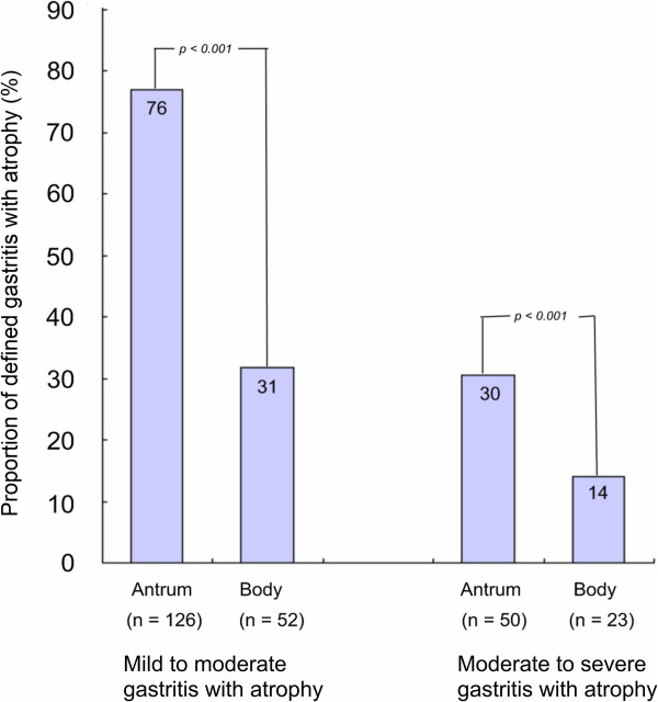
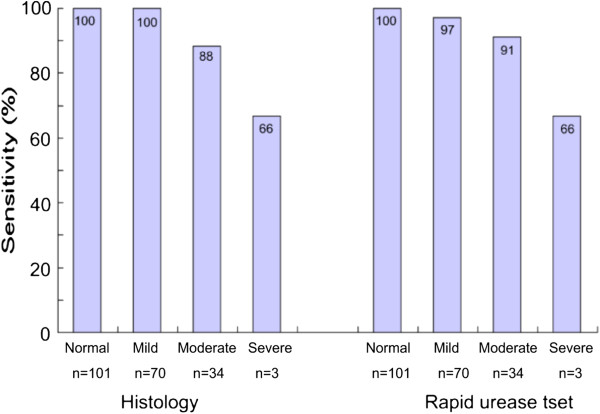
The prevalence rate of H. pylori infection in the 164 patients was 63.4%. Gastritis with atrophy was significantly higher at the antrum than at the body (76% vs. 31%; p<0.001). The sensitivity of biopsy-based test decreased when the degree of gastritis with atrophy increased regardless of biopsy site (for normal, mild, moderate, and severe gastritis with atrophy, the sensitivity of histology Giemsa stain was 100%, 100%, 88%, and 66%, respectively, and 100%, 97%, 91%, and 66%, respectively, for rapid urease test). In moderate to severe antrum or body gastritis with atrophy, additional corpus biopsy resulted in increased sensitivity to 16.67% compare to single antrum biopsy.
In moderate to severe gastritis with atrophy, biopsy-based test should include the corpus for avoiding false negative results.
评估萎缩性胃炎患者 H. pylori 感染的基于活检的检测的最佳部位尚不清楚。本研究旨在评估基于活检的检测在萎缩性胃炎程度方面的部位和敏感性。
纳入 164 例未经调查的消化不良患者。进行基于活检的检测(即培养、组织学吉姆萨染色和快速尿素酶试验)和非侵入性检测(抗 H. pylori IgG)。根据先前的标准定义 H. pylori 感染的金标准。计算胃窦和胃体活检在萎缩性胃炎程度方面的基于活检的检测的敏感性、特异性、阳性预测率和阴性预测率。
164 例患者中 H. pylori 感染的患病率为 63.4%。萎缩性胃炎在胃窦明显高于胃体(76% vs. 31%;p<0.001)。无论活检部位如何,随着萎缩性胃炎程度的增加,基于活检的检测的敏感性都会降低(对于正常、轻度、中度和重度萎缩性胃炎,组织学吉姆萨染色的敏感性分别为 100%、100%、88%和 66%,快速尿素酶试验分别为 100%、97%、91%和 66%)。在中重度胃窦或胃体萎缩性胃炎中,与单独胃窦活检相比,额外的胃体活检可使敏感性提高至 16.67%。
在中重度萎缩性胃炎中,基于活检的检测应包括胃体,以避免假阴性结果。