Department of Orthopaedic Surgery, Saitama Medical University, Saitama, Japan.
J Neuroinflammation. 2013 Jan 3;10:1. doi: 10.1186/1742-2094-10-1.
The physiological function of p38α, which is an isoform of p38 MAPK, has been investigated previously in several studies using pharmacological inhibitors. However, the results regarding whether p38α promotes or inhibits nerve regeneration in vivo have been controversial.
We generated novel p38α mutant mice (sem mice) with a point mutation in the region encoding the p38α substrate-docking-site, which serves as a limited loss-of-function model of p38α. In the present study, we utilized sem mice and wild-type littermates (wt mice) to investigate the physiological role of p38α in nerve regeneration following crush injuries.
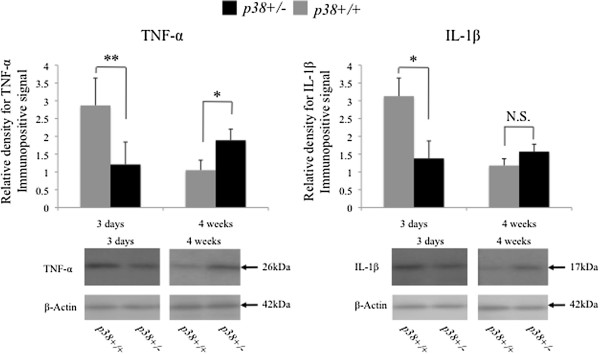
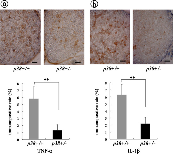
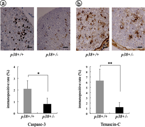
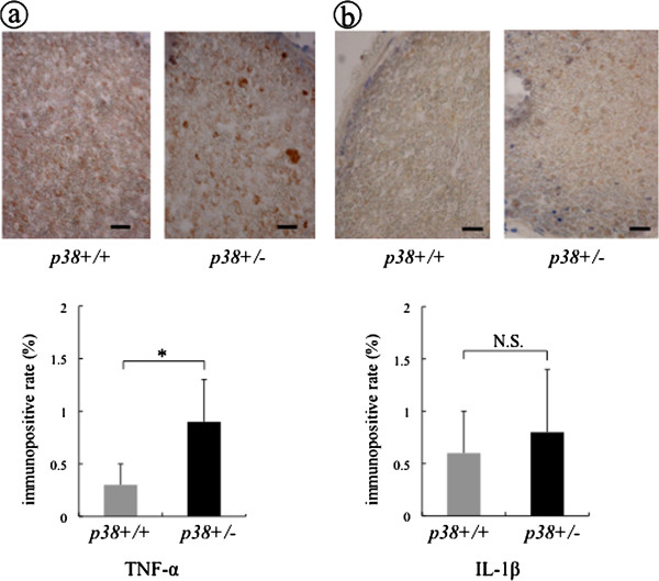
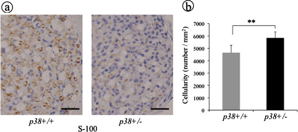
At four weeks after crush injury, the average axon diameter and the average axon area in sem mice were significantly smaller than those in wt mice. The average myelin sheath thickness in sem mice was reduced compared to wt mice, but no significant difference was observed in the G-ratio between the two groups. The sciatic functional index value demonstrated that functional nerve recovery in sem mice following crush injury was delayed, which is consistent with the histological findings. To investigate the underlying mechanisms of these findings, we examined inflammatory responses of the sciatic nerve by immunohistochemistry and western blotting. At an early phase following crush injury, sem mice showed remarkably lower expression of inflammatory cytokines, such as TNF-α and IL-1β, than wt mice. The expression of Caspase-3 and Tenascin-C were also lower in sem mice. Conversely, at a late phase of the response, sem mice showed considerably higher expression of TNF-α and of IL-1β with lower expression of S-100 than wt mice.
This is the first study of the physiological role of p38 MAPK in nerve regeneration that does not rely on the use of pharmacological inhibitors. Our results indicate that p38α insufficiency may cause an inflammatory disorder, resulting in a delay of histological and functional nerve recovery following crush injury. We conclude that p38 MAPK has an important physiological role in nerve regeneration and may be important for controlling both initiation of inflammation and recovery from nerve injury.
p38 MAPK 的同工型 p38α 的生理功能已在使用药理学抑制剂的几项研究中进行了研究。然而,关于 p38α 是否促进或抑制体内神经再生的结果一直存在争议。
我们生成了新型 p38α 突变小鼠(sem 小鼠),该突变小鼠在编码 p38α 底物结合位点的区域中具有点突变,该突变作为 p38α 的有限功能丧失模型。在本研究中,我们利用 sem 小鼠和野生型同窝仔鼠(wt 小鼠)来研究 p38α 在挤压伤后神经再生中的生理作用。
在挤压伤后 4 周时,sem 小鼠的平均轴突直径和平均轴突面积明显小于 wt 小鼠。sem 小鼠的平均髓鞘厚度比 wt 小鼠减少,但两组之间的 G 比值没有差异。坐骨神经功能指数值表明,sem 小鼠在挤压伤后的神经功能恢复延迟,这与组织学发现一致。为了研究这些发现的潜在机制,我们通过免疫组织化学和 Western blot 分析检测了坐骨神经的炎症反应。在挤压伤后早期,sem 小鼠的 TNF-α 和 IL-1β 等炎症细胞因子的表达明显低于 wt 小鼠。sem 小鼠的 Caspase-3 和 Tenascin-C 的表达也较低。相反,在反应的晚期,sem 小鼠的 TNF-α 和 IL-1β 表达较高,而 S-100 表达较低。
这是第一项不依赖于药理学抑制剂研究 p38 MAPK 在神经再生中的生理作用的研究。我们的结果表明,p38α 不足可能导致炎症失调,导致挤压伤后组织学和功能神经恢复延迟。我们得出结论,p38 MAPK 在神经再生中具有重要的生理作用,可能对控制炎症的启动和神经损伤的恢复都很重要。