Department of Anatomy, College of Medicine, The Catholic University of Korea, Seoul, Korea.
PLoS One. 2012;7(12):e52295. doi: 10.1371/journal.pone.0052295. Epub 2012 Dec 17.
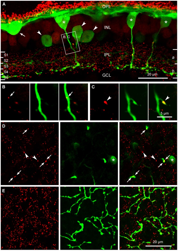
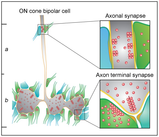
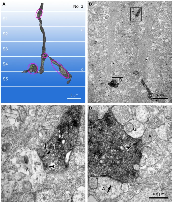
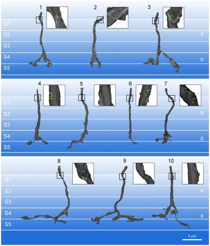
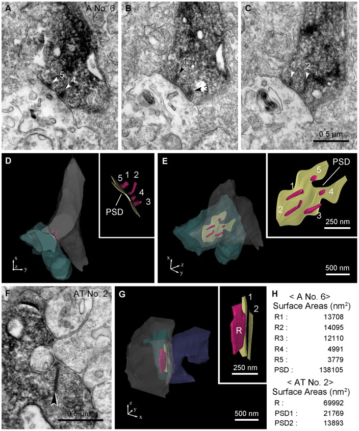
In the mammalian retina, bipolar cells and ganglion cells which stratify in sublamina a of the inner plexiform layer (IPL) show OFF responses to light stimuli while those that stratify in sublamina b show ON responses. This functional relationship between anatomy and physiology is a key principle of retinal organization. However, there are at least three types of retinal neurons, including intrinsically photosensitive retinal ganglion cells (ipRGCs) and dopaminergic amacrine cells, which violate this principle. These cell types have light-driven ON responses, but their dendrites mainly stratify in sublamina a of the IPL, the OFF sublayer. Recent anatomical studies suggested that certain ON cone bipolar cells make axonal or ectopic synapses as they descend through sublamina a, thus providing ON input to cells which stratify in the OFF sublayer. Using immunoelectron microscopy with 3-dimensional reconstruction, we have identified axonal synapses of ON cone bipolar cells in the rabbit retina. Ten calbindin ON cone bipolar axons made en passant ribbon synapses onto amacrine or ganglion dendrites in sublamina a of the IPL. Compared to the ribbon synapses made by bipolar terminals, these axonal ribbon synapses were characterized by a broad postsynaptic element that appeared as a monad and by the presence of multiple short synaptic ribbons. These findings confirm that certain ON cone bipolar cells can provide ON input to amacrine and ganglion cells whose dendrites stratify in the OFF sublayer via axonal synapses. The monadic synapse with multiple ribbons may be a diagnostic feature of the ON cone bipolar axonal synapse in sublamina a. The presence of multiple ribbons and a broad postsynaptic density suggest these structures may be very efficient synapses. We also identified axonal inputs to ipRGCs with the architecture described above.
在哺乳动物的视网膜中,位于内丛状层(IPL)亚层 a 的双极细胞和节细胞对光刺激表现出 OFF 反应,而位于亚层 b 的双极细胞和节细胞则表现出 ON 反应。这种解剖结构和生理功能之间的关系是视网膜组织的一个关键原则。然而,至少有三种类型的视网膜神经元,包括内在光敏视网膜节细胞(ipRGCs)和多巴胺能无长突细胞,违反了这一原则。这些细胞类型具有光驱动的 ON 反应,但它们的树突主要位于 IPL 的亚层 a,即 OFF 亚层。最近的解剖学研究表明,某些 ON 锥形双极细胞在穿过亚层 a 下降的过程中形成轴突或异位突触,从而为位于 OFF 亚层的细胞提供 ON 输入。通过使用三维重建的免疫电子显微镜,我们在兔视网膜中鉴定出了 ON 锥形双极细胞的轴突突触。10 个钙结合蛋白 ON 锥形双极细胞轴突在 IPL 的亚层 a 中形成了与无长突或节细胞树突的瞬间连接性带状突触。与双极细胞末端形成的带状突触相比,这些轴突带状突触的特征是具有宽的后突触元件,看起来像单体,并且存在多个短的突触小泡。这些发现证实,某些 ON 锥形双极细胞可以通过轴突突触向位于 OFF 亚层的无长突和节细胞提供 ON 输入。具有多个小泡的单体突触可能是亚层 a 中 ON 锥形双极细胞轴突突触的一个诊断特征。多个小泡和宽的后突触密度表明这些结构可能是非常有效的突触。我们还鉴定出了具有上述结构的 ipRGCs 的轴突输入。