• 文献检索
  • 文档翻译
  • 深度研究
  • 学术资讯
  • Suppr Zotero 插件Zotero 插件
  • 邀请有礼
  • 套餐&价格
  • 历史记录
应用&插件
Suppr Zotero 插件Zotero 插件浏览器插件Mac 客户端Windows 客户端微信小程序
定价
高级版会员购买积分包购买API积分包
服务
文献检索文档翻译深度研究API 文档MCP 服务
关于我们
关于 Suppr公司介绍联系我们用户协议隐私条款
关注我们

Suppr 超能文献

核心技术专利:CN118964589B侵权必究
粤ICP备2023148730 号-1Suppr @ 2026

文献检索

告别复杂PubMed语法,用中文像聊天一样搜索,搜遍4000万医学文献。AI智能推荐,让科研检索更轻松。

立即免费搜索

文件翻译

保留排版,准确专业,支持PDF/Word/PPT等文件格式,支持 12+语言互译。

免费翻译文档

深度研究

AI帮你快速写综述,25分钟生成高质量综述,智能提取关键信息,辅助科研写作。

立即免费体验

应用靶向抗 CD31 抗体的共聚焦激光内镜对人结直肠癌微血管进行形态计量评估。

Confocal laser endomicroscopy for the morphometric evaluation of microvessels in human colorectal cancer using targeted anti-CD31 antibodies.

机构信息

Research Center of Gastroenterology and Hepatology, University of Medicine and Pharmacy of Craiova, Craiova, Romania.

出版信息

PLoS One. 2012;7(12):e52815. doi: 10.1371/journal.pone.0052815. Epub 2012 Dec 28.

DOI:10.1371/journal.pone.0052815
PMID:23285192
原文链接:https://pmc.ncbi.nlm.nih.gov/articles/PMC3532115/
Abstract

INTRODUCTION

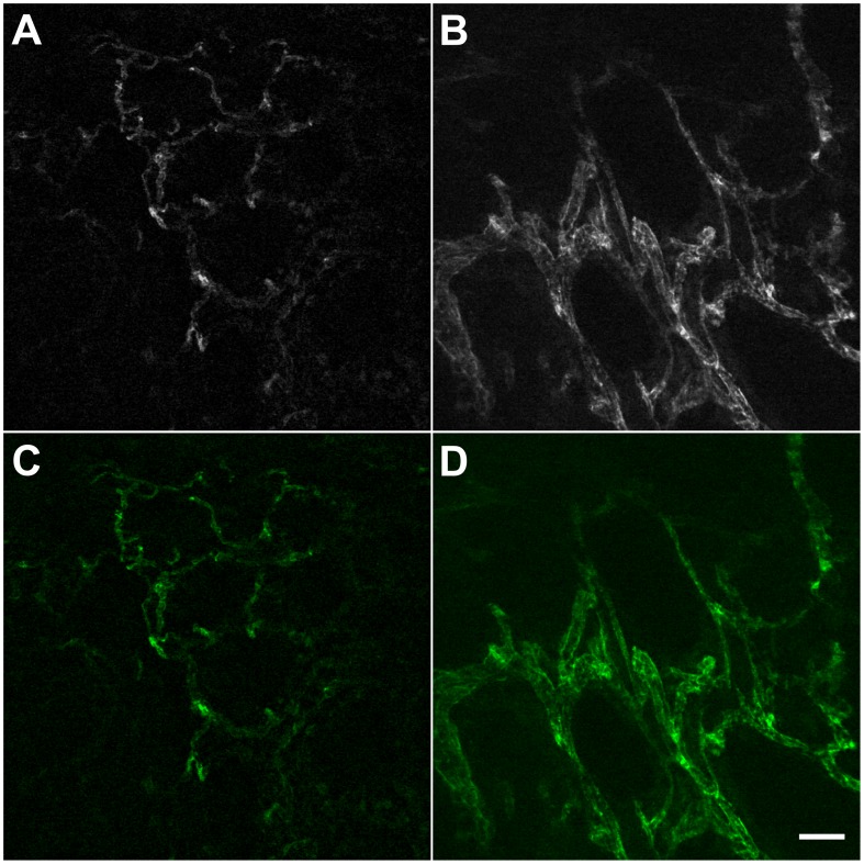
Numerous anti-angiogenic agents are currently developed to limit tumor growth and metastasis. While these drugs offer hope for cancer patients, their transient effect on tumor vasculature is difficult to assess in clinical settings. Confocal laser endomicroscopy (CLE) is a novel endoscopic imaging technology that enables histological examination of the gastrointestinal mucosa. The aim of the present study was to evaluate the feasibility of using CLE to image the vascular network in fresh biopsies of human colorectal tissue. For this purpose we have imaged normal and malignant biopsy tissue samples and compared the vascular network parameters obtained with CLE with established histopathology techniques.

MATERIALS AND METHODS

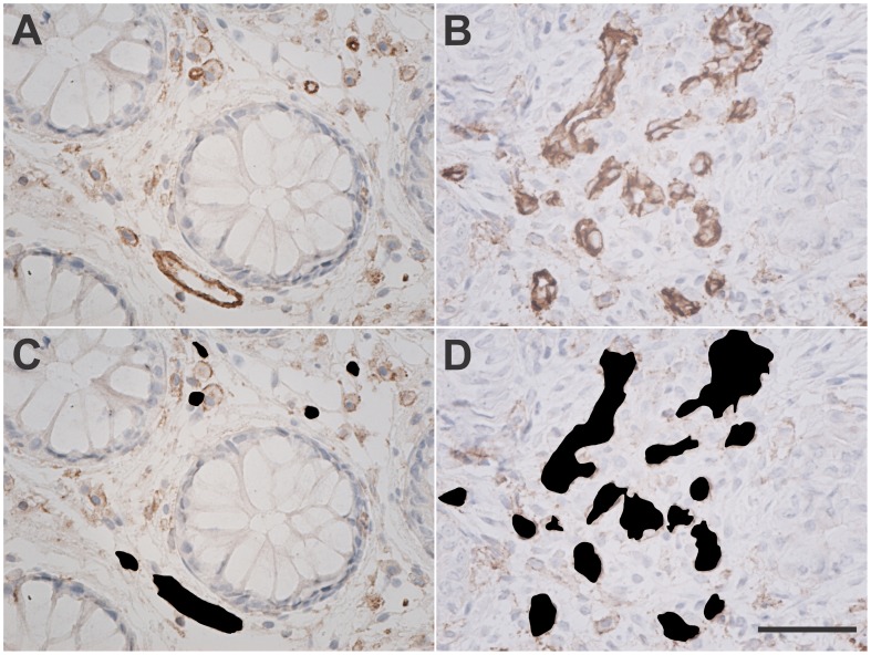
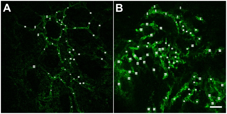
Fresh non-fixed biopsy samples of both normal and malignant colorectal mucosa were stained with fluorescently labeled anti-CD31 antibodies and imaged by CLE using a dedicated endomicroscopy system. Corresponding biopsy samples underwent immunohistochemical staining for CD31, assessing the microvessel density (MVD) and vascular areas for comparison with CLE data, which were measured offline using specific software.

RESULTS

The vessels were imaged by CLE in both normal and tumor samples. The average diameter of normal vessels was 8.5±0.9 µm whereas in tumor samples it was 13.5±0.7 µm (p = 0.0049). Vascular density was 188.7±24.9 vessels/mm(2) in the normal tissue vs. 242.4±16.1 vessels/mm(2) in the colorectal cancer samples (p = 0.1201). In the immunohistochemistry samples, the MVD was 211.2±42.9/mm(2) and 351.3±39.6/mm(2) for normal and malignant mucosa, respectively. The vascular area was 2.9±0.5% of total tissue area for the normal mucosa and 8.5±2.1% for primary colorectal cancer tissue.

CONCLUSION

Selective imaging of blood vessels with CLE is feasible in normal and tumor colorectal tissue by using fluorescently labeled antibodies targeted against an endothelial marker. The method could be translated into the clinical setting for monitoring of anti-angiogenic therapy.

摘要

简介

目前有许多抗血管生成药物被开发出来,以限制肿瘤的生长和转移。虽然这些药物为癌症患者带来了希望,但它们对肿瘤血管系统的短暂作用在临床环境中很难评估。共聚焦激光内镜检查(CLE)是一种新型的内镜成像技术,可对胃肠道黏膜进行组织学检查。本研究旨在评估使用 CLE 对人结直肠组织新鲜活检标本中的血管网络进行成像的可行性。为此,我们对正常和恶性活检组织样本进行了成像,并将通过 CLE 获得的血管网络参数与已建立的组织病理学技术进行了比较。

材料和方法

用荧光标记的抗 CD31 抗体对新鲜非固定的结直肠黏膜活检样本进行染色,并使用专用的内镜检查系统进行 CLE 成像。对相应的活检样本进行 CD31 的免疫组织化学染色,评估微血管密度(MVD)和血管面积,与 CLE 数据进行比较,后者使用特定软件在线下进行测量。

结果

CLE 可以在正常和肿瘤样本中对血管进行成像。正常血管的平均直径为 8.5±0.9 µm,而在肿瘤样本中为 13.5±0.7 µm(p=0.0049)。正常组织的血管密度为 188.7±24.9 条/mm(2),结直肠癌样本为 242.4±16.1 条/mm(2)(p=0.1201)。在免疫组化样本中,正常和恶性黏膜的 MVD 分别为 211.2±42.9/mm(2)和 351.3±39.6/mm(2)。正常黏膜的血管面积占总组织面积的 2.9±0.5%,原发性结直肠癌组织的血管面积占 8.5±2.1%。

结论

通过使用针对内皮标志物的荧光标记抗体,CLE 可以在正常和肿瘤结直肠组织中选择性地对血管进行成像。该方法可转化为临床环境,用于监测抗血管生成治疗。

https://cdn.ncbi.nlm.nih.gov/pmc/blobs/6db3/3532115/bfe232d2011b/pone.0052815.g003.jpg
https://cdn.ncbi.nlm.nih.gov/pmc/blobs/6db3/3532115/20665b4d489f/pone.0052815.g001.jpg
https://cdn.ncbi.nlm.nih.gov/pmc/blobs/6db3/3532115/ada09cd293b7/pone.0052815.g002.jpg
https://cdn.ncbi.nlm.nih.gov/pmc/blobs/6db3/3532115/bfe232d2011b/pone.0052815.g003.jpg
https://cdn.ncbi.nlm.nih.gov/pmc/blobs/6db3/3532115/20665b4d489f/pone.0052815.g001.jpg
https://cdn.ncbi.nlm.nih.gov/pmc/blobs/6db3/3532115/ada09cd293b7/pone.0052815.g002.jpg
https://cdn.ncbi.nlm.nih.gov/pmc/blobs/6db3/3532115/bfe232d2011b/pone.0052815.g003.jpg

相似文献

1
Confocal laser endomicroscopy for the morphometric evaluation of microvessels in human colorectal cancer using targeted anti-CD31 antibodies.应用靶向抗 CD31 抗体的共聚焦激光内镜对人结直肠癌微血管进行形态计量评估。
PLoS One. 2012;7(12):e52815. doi: 10.1371/journal.pone.0052815. Epub 2012 Dec 28.
2
Evaluation of new morphometric parameters of neoangiogenesis in human colorectal cancer using confocal laser endomicroscopy (CLE) and targeted panendothelial markers.使用共聚焦激光内镜显微镜(CLE)和靶向全内皮标记物评估人类结直肠癌新生血管生成的新形态学参数。
PLoS One. 2014 Mar 10;9(3):e91084. doi: 10.1371/journal.pone.0091084. eCollection 2014.
3
Tumor neoangiogenesis detection by confocal laser endomicroscopy and anti-CD105 antibody: Pilot study.共聚焦激光内镜联合抗CD105抗体检测肿瘤新生血管:初步研究
World J Gastrointest Oncol. 2015 Nov 15;7(11):361-8. doi: 10.4251/wjgo.v7.i11.361.
4
In vivo assessment of tumour angiogenesis in colorectal cancer: the role of confocal laser endomicroscopy.结直肠癌中肿瘤血管生成的体内评估:共聚焦激光内镜显微镜的作用
Colorectal Dis. 2016 Feb;18(2):O66-73. doi: 10.1111/codi.13222.
5
In vivo molecular imaging of colorectal cancer with confocal endomicroscopy by targeting epidermal growth factor receptor.通过靶向表皮生长因子受体进行共聚焦内镜术对结直肠癌的体内分子成像。
Gastroenterology. 2010 Feb;138(2):435-46. doi: 10.1053/j.gastro.2009.10.032. Epub 2009 Oct 21.
6
Computer Aided Diagnosis for Confocal Laser Endomicroscopy in Advanced Colorectal Adenocarcinoma.晚期结直肠癌共聚焦激光内镜检查的计算机辅助诊断
PLoS One. 2016 May 4;11(5):e0154863. doi: 10.1371/journal.pone.0154863. eCollection 2016.
7
VEGFR2 heterogeneity and response to anti-angiogenic low dose metronomic cyclophosphamide treatment.VEGFR2 异质性与抗血管生成低剂量节拍式环磷酰胺治疗反应。
BMC Cancer. 2010 Dec 15;10:683. doi: 10.1186/1471-2407-10-683.
8
[Confocal laser endomicroscopy is a new endoscopic technique for diagnosing colorectal neoplasia and inflammatory bowel disease].共聚焦激光内镜检查术是一种用于诊断结直肠肿瘤和炎症性肠病的新型内镜技术。
Ugeskr Laeger. 2014 Sep 29;176(40).
9
Confocal laser endomicroscopy: a new gold standard for the assessment of mucosal healing in ulcerative colitis.共聚焦激光内镜检查术:评估溃疡性结肠炎黏膜愈合的一项新的金标准。
J Gastroenterol Hepatol. 2015 Mar;30 Suppl 1:85-92. doi: 10.1111/jgh.12748.
10
Technology insight: confocal laser endoscopy for in vivo diagnosis of colorectal cancer.技术洞察:用于结直肠癌体内诊断的共聚焦激光内镜检查
Nat Clin Pract Oncol. 2007 Aug;4(8):480-90. doi: 10.1038/ncponc0881.

引用本文的文献

1
Current trends in the characterization and monitoring of vascular response to cancer therapy.当前癌症治疗中血管反应的特征和监测趋势。
Cancer Imaging. 2024 Oct 23;24(1):143. doi: 10.1186/s40644-024-00767-8.
2
Molecular Profiling of EGFR Status to Identify Skin Toxicity in Colorectal Cancer: A Clinicopathological Review.通过表皮生长因子受体(EGFR)状态的分子分析来识别结直肠癌中的皮肤毒性:一项临床病理综述
Curr Health Sci J. 2019 Apr-Jun;45(2):127-133. doi: 10.12865/CHSJ.45.02.01. Epub 2019 Jun 30.
3
Vascular architecture in the bacteriogenic light organ of (Cephalopoda: Sepiolidae).

本文引用的文献

1
Molecular imaging of epidermal growth factor-receptor and survivin in vivo in porcine esophageal and gastric mucosae using probe-based confocal laser-induced endomicroscopy: proof of concept.基于探针的共聚焦激光诱导内镜在猪食管和胃黏膜中对表皮生长因子受体和生存素的分子成像:概念验证。
J Physiol Pharmacol. 2012 Jun;63(3):303-7.
2
Miami classification for probe-based confocal laser endomicroscopy.基于探针的共聚焦激光内镜的迈阿密分类。
Endoscopy. 2011 Oct;43(10):882-91. doi: 10.1055/s-0030-1256632. Epub 2011 Aug 4.
3
Confocal laser endomicroscopy and immunoendoscopy for real-time assessment of vascularization in gastrointestinal malignancies.
(头足纲:乌贼科)产菌性发光器官的血管结构。
Invertebr Biol. 2018 Sep;137(3):240-249. doi: 10.1111/ivb.12223. Epub 2018 Sep 7.
4
Endomicroscopy with Fluorescent CD105 Antibodies for "In Vivo" Imaging of Colorectal Cancer Angiogenesis.使用荧光CD105抗体的内镜显微镜检查用于结直肠癌血管生成的“体内”成像
Curr Health Sci J. 2015 Jul-Sep;41(3):288-292. doi: 10.12865/CHSJ.41.03.17. Epub 2015 Mar 15.
5
Assessing tumor angiogenesis in colorectal cancer by quantitative contrast-enhanced endoscopic ultrasound and molecular and immunohistochemical analysis.通过定量对比增强内镜超声以及分子和免疫组织化学分析评估结直肠癌中的肿瘤血管生成。
Endosc Ultrasound. 2018 May-Jun;7(3):175-183. doi: 10.4103/eus.eus_7_17.
6
Detection of colonic dysplasia in patients with ulcerative colitis using a targeted fluorescent peptide and confocal laser endomicroscopy: A pilot study.使用靶向荧光肽和共聚焦激光内镜显微镜检测溃疡性结肠炎患者的结肠发育异常:一项初步研究。
PLoS One. 2017 Jun 30;12(6):e0180509. doi: 10.1371/journal.pone.0180509. eCollection 2017.
7
Vector-mediated Tum-5 expression in neovascular endothelial cells for treating hepatocellular carcinoma.通过载体介导在新生血管内皮细胞中表达Tum-5用于治疗肝细胞癌。
Exp Ther Med. 2017 Apr;13(4):1521-1525. doi: 10.3892/etm.2017.4127. Epub 2017 Feb 15.
8
Development and Validation of a Histological Method to Measure Microvessel Density in Whole-Slide Images of Cancer Tissue.一种用于测量癌组织全切片图像中微血管密度的组织学方法的开发与验证
PLoS One. 2016 Sep 1;11(9):e0161496. doi: 10.1371/journal.pone.0161496. eCollection 2016.
9
Computer Aided Diagnosis for Confocal Laser Endomicroscopy in Advanced Colorectal Adenocarcinoma.晚期结直肠癌共聚焦激光内镜检查的计算机辅助诊断
PLoS One. 2016 May 4;11(5):e0154863. doi: 10.1371/journal.pone.0154863. eCollection 2016.
10
Confocal Laser Endomicroscopy in Gastrointestinal and Pancreatobiliary Diseases: A Systematic Review and Meta-Analysis.共聚焦激光内镜检查在胃肠和胰胆疾病中的应用:一项系统评价和Meta分析
Biomed Res Int. 2016;2016:4638683. doi: 10.1155/2016/4638683. Epub 2016 Feb 17.
共聚焦激光内镜检查和免疫内镜检查实时评估胃肠道恶性肿瘤的血管生成。
World J Gastroenterol. 2011 Jan 7;17(1):21-7. doi: 10.3748/wjg.v17.i1.21.
4
Narrow-banding images and structures of microvessels of colonic lesions.窄带成像技术与结直肠病变黏膜下微血管的显影
Dig Dis Sci. 2011 Jun;56(6):1811-7. doi: 10.1007/s10620-010-1519-4. Epub 2010 Dec 28.
5
Vascular endothelial platelet endothelial cell adhesion molecule 1 (PECAM-1) regulates advanced metastatic progression.血管内皮细胞血小板内皮细胞黏附分子 1(PECAM-1)调节晚期转移进展。
Proc Natl Acad Sci U S A. 2010 Oct 26;107(43):18616-21. doi: 10.1073/pnas.1004654107. Epub 2010 Oct 6.
6
Molecular imaging of VEGF in gastrointestinal cancer in vivo using confocal laser endomicroscopy.应用共聚焦激光内镜术对胃肠道癌中 VEGF 的分子成像。
Gut. 2010 Aug;59(8):1046-55. doi: 10.1136/gut.2009.202986.
7
The spatial arrangement of the human large intestinal wall blood circulation.人体大肠壁血液循环的空间排列。
J Anat. 2010 Mar;216(3):335-43. doi: 10.1111/j.1469-7580.2009.01199.x.
8
Advances of endomicroscopy for gastrointestinal physiology and diseases.内窥式显微镜在胃肠道生理学和疾病中的应用进展。
Am J Physiol Gastrointest Liver Physiol. 2010 Jun;298(6):G797-806. doi: 10.1152/ajpgi.00027.2010. Epub 2010 Feb 25.
9
In vivo molecular imaging of colorectal cancer with confocal endomicroscopy by targeting epidermal growth factor receptor.通过靶向表皮生长因子受体进行共聚焦内镜术对结直肠癌的体内分子成像。
Gastroenterology. 2010 Feb;138(2):435-46. doi: 10.1053/j.gastro.2009.10.032. Epub 2009 Oct 21.
10
In vivo diagnosis and classification of colorectal neoplasia by chromoendoscopy-guided confocal laser endomicroscopy.内窥激光共聚焦显微镜下靶向染色指导的结直肠肿瘤的体内诊断和分类。
Clin Gastroenterol Hepatol. 2010 Apr;8(4):371-8. doi: 10.1016/j.cgh.2009.08.006. Epub 2009 Aug 13.