De Palma Giovanni Domenico, Colavita Irene, Zambrano Gerardo, Giglio Mariano Cesare, Maione Francesco, Luglio Gaetano, Sarnelli Giovanni, Rispo Antonio, Schettino Pietro, D'Armiento Francesco Paolo, De Palma Fatima Domenica Elisa, D'Argenio Valeria, Salvatore Francesco
Department of Clinical Medicine and Surgery, University of Naples Federico II, Naples, Italy.
CEINGE-Biotecnologie Avanzate, Naples, Italy.
PLoS One. 2017 Jun 30;12(6):e0180509. doi: 10.1371/journal.pone.0180509. eCollection 2017.
Targeted molecular probes have been used to detect sporadic colonic dysplasia during confocal laser endomicroscopy (CLE) with promising results. This is a feasibility pilot study aiming to assess the potential role of CLE combined with a fluorescent-labeled peptide to stain and detect dysplasia associated with Ulcerative Colitis.
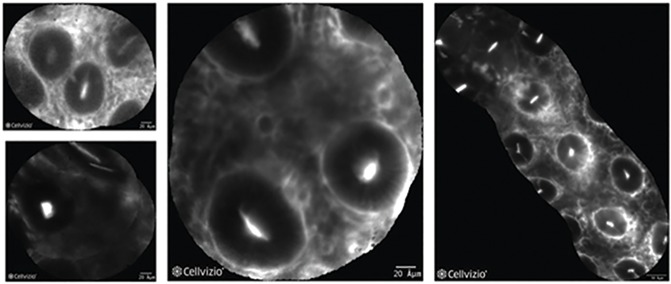
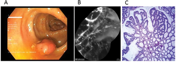
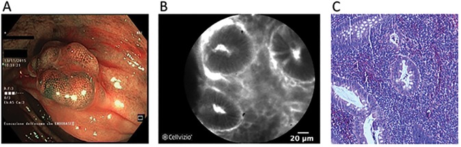
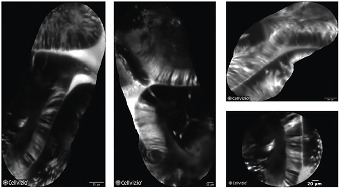
A phage-derived heptapeptide with predicted high binding affinity for dysplastic tissue, was synthesized and labeled with fluorescein. Eleven lesions with suspected dysplasia at endoscopy were excised from nine patients with long-standing ulcerative colitis. Specimens were sprayed with the peptide and examined by CLE. The CLE images were then compared to the corresponding histological sections.
At definitive histology, 4 lesions were diagnosed as inflammatory polyps, 6 as dysplastic lesions and one as invasive cancer. In inflammatory polyps, the fluorescence signal came from peri-cryptal spaces and crypt lumen due to passive accumulation of the peptide in these areas. Dysplasia was associated with active binding of the peptide to dysplastic colonocytes.
Ex vivo staining of ulcerative colitis-associated dysplasia using a fluorescent labeled molecular probe and CLE is feasible. In vivo studies on larger populations are required to evaluate the safety and the effective contribution of molecular probes in cancer surveillance of ulcerative colitis.
靶向分子探针已被用于在共聚焦激光内镜检查(CLE)期间检测散发性结肠发育异常,取得了有前景的结果。这是一项可行性初步研究,旨在评估CLE联合荧光标记肽对溃疡性结肠炎相关发育异常进行染色和检测的潜在作用。
合成一种对发育异常组织具有预测高结合亲和力的噬菌体衍生七肽,并用荧光素标记。从9例长期溃疡性结肠炎患者中切除11个在内镜检查时疑似发育异常的病变。将标本用该肽喷洒并通过CLE检查。然后将CLE图像与相应的组织学切片进行比较。
在最终组织学检查中,4个病变被诊断为炎性息肉,6个为发育异常病变,1个为浸润性癌。在炎性息肉中,荧光信号来自隐窝周围间隙和隐窝腔,这是由于该肽在这些区域的被动积聚。发育异常与该肽与发育异常结肠细胞的主动结合有关。
使用荧光标记分子探针和CLE对溃疡性结肠炎相关发育异常进行离体染色是可行的。需要对更大规模人群进行体内研究,以评估分子探针在溃疡性结肠炎癌症监测中的安全性和有效作用。