Perkins Guy A, Scott Ray, Perez Alex, Ellisman Mark H, Johnson Jerry E, Fox Donald A
National Center for Microscopy and Imaging Research, University of California San Diego, La Jolla, CA, USA.
Mol Vis. 2012;18:3029-48. Epub 2012 Dec 20.
Postnatal lead exposure produces rod-selective and Bax-mediated apoptosis, decreased scotopic electroretinograms (ERGs), and scotopic and mesopic vision deficits in humans and/or experimental animals. Rod, but not cone, inner segment mitochondria were considered the primary site of action. However, photoreceptor synaptic mitochondria were not examined. Thus, our experiments investigated the structural and functional effects of environmentally relevant postnatal lead exposure on rod spherule and cone pedicle mitochondria and whether Bcl-xL overexpression provided neuroprotection.
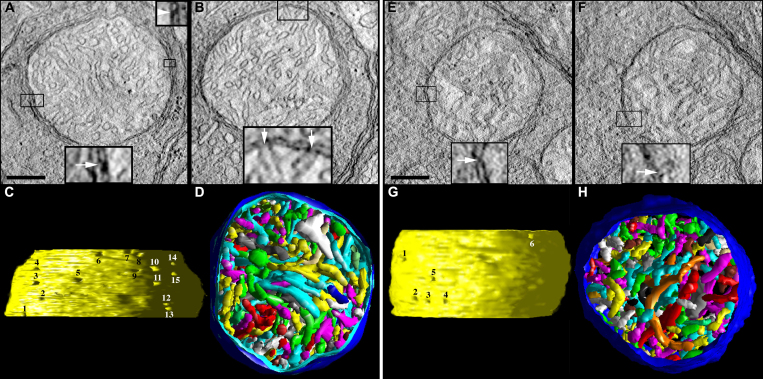
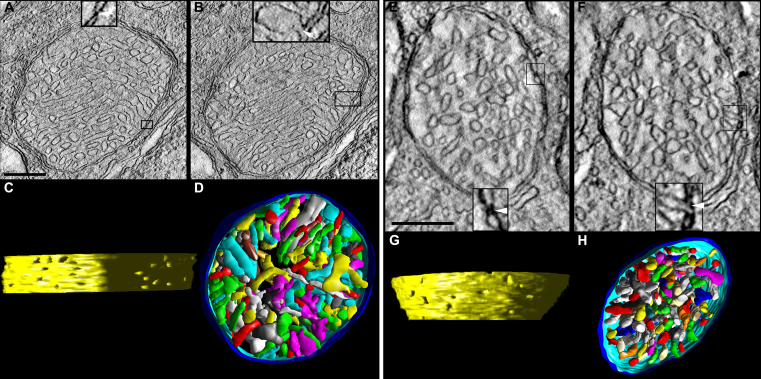
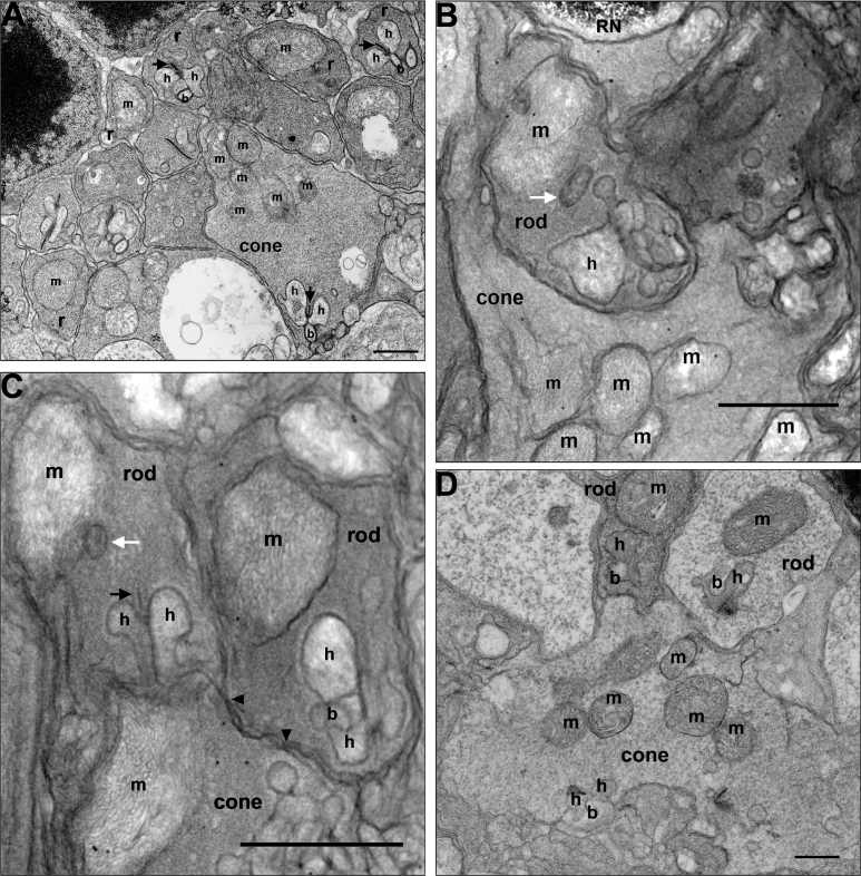
C57BL/6N mice pups were exposed to lead only during lactation via dams drinking water containing lead acetate. The blood [Pb] at weaning was 20.6±4.7 µg/dl, which decreased to the control value by 2 months. To assess synaptic mitochondrial structural differences and vulnerability to lead exposure, wild-type and transgenic mice overexpressing Bcl-xL in photoreceptors were used. Electron microscopy, three-dimensional electron tomography, and retinal and photoreceptor synaptic terminal oxygen consumption (QO(2)) studies were conducted in adult control, Bcl-xL, lead, and Bcl-xL/lead mice.
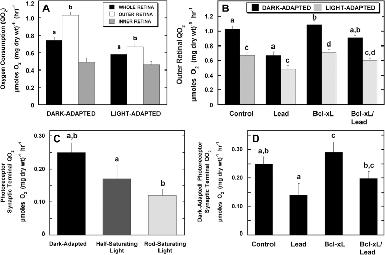
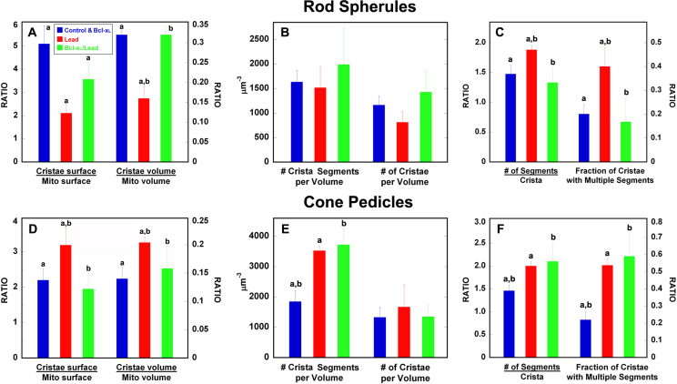
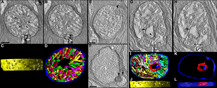
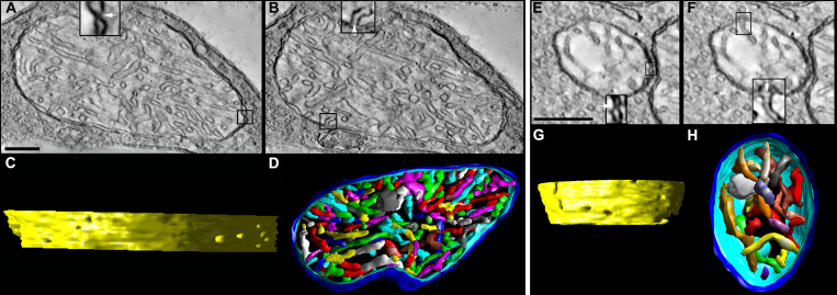
The spherule and pedicle mitochondria in lead-treated mice were swollen, and the cristae structure was markedly changed. In the lead-treated mice, the mitochondrial cristae surface area and volume (abundance: measure correlated with ATP (ATP) synthesis) were decreased in the spherules and increased in the pedicles. Pedicles also had an increased number of crista segments per volume. In the lead-treated mice, the number of segments/crista and fraction of cristae with multiple segments (branching) similarly increased in spherule and pedicle mitochondria. Lead-induced remodeling of spherule mitochondria produced smaller cristae with more branching, whereas pedicle mitochondria had larger cristae with more branching and increased crista junction (CJ) diameter. Lead decreased dark- and light-adapted photoreceptor and dark-adapted photoreceptor synaptic terminal QO(2). Bcl-xL partially blocked many of the lead-induced alterations relative to controls. However, spherules still had partially decreased abundance, whereas pedicles still had increased branching, increased crista segments per volume, and increased crista junction diameter. Moreover, photoreceptor and synaptic QO(2) were only partially recovered.
These findings reveal cellular and compartmental specific differences in the structure and vulnerability of rod and cone inner segment and synaptic mitochondria to postnatal lead exposure. Spherule and pedicle mitochondria in lead-exposed mice displayed complex and distinguishing patterns of cristae and matrix damage and remodeling consistent with studies showing that synaptic mitochondria are more sensitive to Ca(2+) overload, oxidative stress, and ATP loss than non-synaptic mitochondria. The lead-induced decreases in QO(2) likely resulted from the decreased spherule cristae abundance and smaller cristae, perhaps due to Bax-mediated effects as they occurred in apoptotic rod inner segments. The increase in pedicle cristae abundance and CJ diameter could have resulted from increased Drp1-mediated fission, as small mitochondrial fragments were observed. The mechanisms of Bcl-xL-mediated remodeling might occur via interaction with formation of CJ protein 1 (Fcj1), whereas the partial protection of synaptic QO(2) might result from the enhanced efficiency of energy metabolism via Bcl-xL's direct interaction with the F1F0 ATP synthase and/or regulation of cellular redox status. These lead-induced alterations in photoreceptor synaptic terminal mitochondria likely underlie the persistent scotopic and mesopic deficits in lead-exposed children, workers, and experimental animals. Our findings stress the clinical and scientific importance of examining synaptic dysfunction following injury or disease during development, and developing therapeutic treatments that prevent synaptic degeneration in retinal and neurodegenerative disorders even when apoptosis is blocked.
出生后铅暴露会导致视杆细胞选择性和 Bax 介导的细胞凋亡、暗视视网膜电图(ERG)降低,以及人类和/或实验动物出现暗视和中间视觉缺陷。视杆细胞而非视锥细胞的内节线粒体被认为是主要作用部位。然而,尚未对视光感受器突触线粒体进行研究。因此,我们的实验研究了与环境相关的出生后铅暴露对视杆小球和视锥小足线粒体的结构和功能影响,以及 Bcl-xL 的过表达是否能提供神经保护作用。
C57BL/6N 小鼠幼崽仅在哺乳期通过其母鼠饮用含醋酸铅的水来接触铅。断奶时血液中的[Pb]为 20.6±4.7 µg/dl,到 2 个月时降至对照值。为评估突触线粒体的结构差异以及对铅暴露的易感性,使用了在光感受器中过表达 Bcl-xL 的野生型和转基因小鼠。对成年对照小鼠、Bcl-xL 小鼠、铅暴露小鼠和 Bcl-xL/铅暴露小鼠进行了电子显微镜检查、三维电子断层扫描以及视网膜和光感受器突触终末的氧消耗(QO₂)研究。
铅处理小鼠的小球和小足线粒体肿胀,嵴结构明显改变。在铅处理小鼠中