Ko Sheung-Fat, Sheu Jiunn-Jye, Lee Chen-Chang, Huang Chung-Cheng, Lee Fan-Yen, Ng Shu-Hang, Lee Yi-Wei, Yip Hon-Kan, Chen Min-Chi
Department of Radiology, Kaohsiung Chang Gung Memorial Hospital and Chang Gung University College of Medicine, 123 Ta-Pei Road, Niao-Sung District, Kaohsiung 833, Taiwan.
ScientificWorldJournal. 2012;2012:192150. doi: 10.1100/2012/192150. Epub 2012 Dec 11.
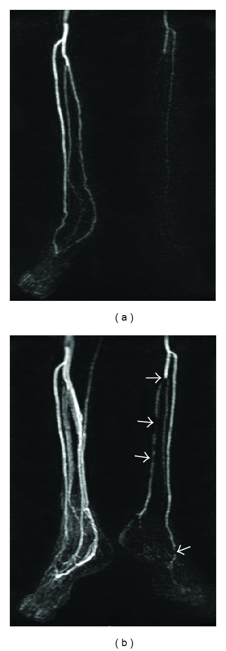
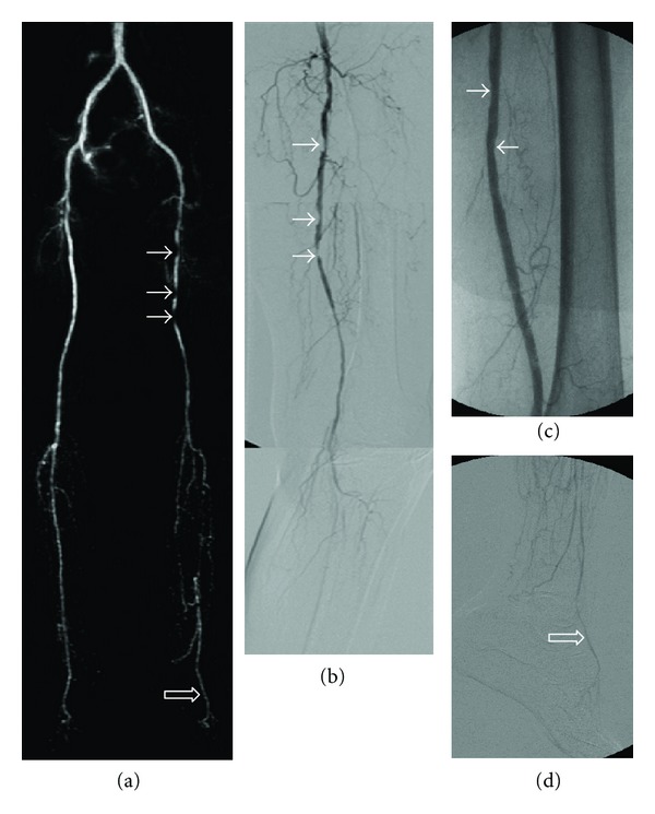
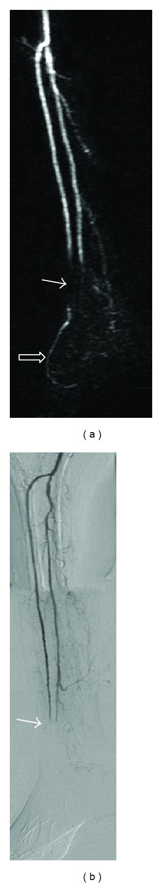
The entire vascular tree of 58 lower extremities with high-grade critical limb ischemia (CLI) was assessed with three-station time resolved imaging of contrast kinetics (TRICKS) magnetic resonance angiography (T-MRA) and correlated with digital subtraction angiography (DSA) examinations and Trans-Atlantic Inter-Society Consensus II (TASC II) guidelines. Kappa (κ) statistics were utilized to evaluate the agreement of stenosis scores (5-point scale; 0 normal to 4 occlusion) based on T-MRA and DSA. With DSA as the standard, significant stenosis instances (stenosis score ≥2) among vascular segments were compared. The κ-statistics of image quality (4-point scale; 1 nondiagnostic to 4 excellent) of T-MRA and TASC II classification assessed by a radiologist and a vascular surgeon were also evaluated. Among 870 vascular segments, excellent agreement was observed between T-MRA and DSA (mean κ = 0.883) in revealing stenosis (mean stenosis score, 2.1 ± 1.3 versus 2.0 ± 1.3). T-MRA harbored overall high sensitivity (99.5%), specificity (93.6%), positive predictive value (95.4%), negative predictive value (99.6%), and accuracy (97.7%) in depicting significant stenosis. Excellent interobserver agreement (mean κ = 0.818) of superb image quality (mean score = 3.5-3.6) of T-MRA and outstanding agreement of TASC II classification of aortoiliac and femoral-popliteal lesions (κ = 0.912-0.917) between two raters further verified the clinical feasibility of T-MRA for treatment planning.
采用三站对比剂动力学时间分辨成像(TRICKS)磁共振血管造影(T-MRA)对58例下肢重度严重肢体缺血(CLI)患者的整个血管树进行评估,并与数字减影血管造影(DSA)检查及《跨大西洋两岸多学会共识II(TASC II)》指南进行对照。利用Kappa(κ)统计量评估基于T-MRA和DSA的狭窄评分(5分制;0为正常至4为闭塞)的一致性。以DSA为标准,比较血管节段中的显著狭窄情况(狭窄评分≥2)。还评估了放射科医生和血管外科医生对T-MRA图像质量(4分制;1为非诊断性至4为优秀)和TASC II分类的κ统计量。在870个血管节段中,T-MRA和DSA在显示狭窄方面具有高度一致性(平均κ = 0.883)(平均狭窄评分分别为2.1±1.3和2.0±1.3)。T-MRA在描绘显著狭窄方面总体具有高敏感性(99.5%)、特异性(93.6%)、阳性预测值(95.4%)、阴性预测值(99.6%)和准确性(97.7%)。两位评估者对T-MRA的出色图像质量(平均评分 = 3.5 - 3.6)具有高度的观察者间一致性(平均κ = 0.818),并且对主-髂动脉和股-腘动脉病变的TASC II分类具有出色的一致性(κ = 0.912 - 0.917),这进一步证实了T-MRA用于治疗规划的临床可行性。