Hoffmann Max, Schwarz Ulrich S
BioQuant, Heidelberg University, Im Neuenheimer Feld 267, 69120 Heidelberg, Germany.
BMC Syst Biol. 2013 Jan 12;7:2. doi: 10.1186/1752-0509-7-2.
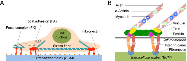
Focal adhesions are integrin-based cell-matrix contacts that transduce and integrate mechanical and biochemical cues from the environment. They develop from smaller and more numerous focal complexes under the influence of mechanical force and are key elements for many physiological and disease-related processes, including wound healing and metastasis. More than 150 different proteins localize to focal adhesions and have been systematically classified in the adhesome project (http://www.adhesome.org). First RNAi-screens have been performed for focal adhesions and the effect of knockdown of many of these components on the number, size, shape and location of focal adhesions has been reported.
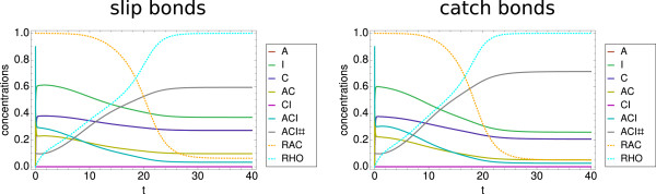
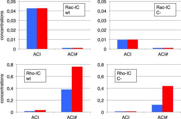
We have developed a kinetic model for RNA interference of focal adhesions which represents some of its main elements: a spatially layered structure, signaling through the small GTPases Rac and Rho, and maturation from focal complexes to focal adhesions under force. The response to force is described by two complementary scenarios corresponding to slip and catch bond behavior, respectively. Using estimated and literature values for the model parameters, three time scales of the dynamics of RNAi-influenced focal adhesions are identified: a sub-minute time scale for the assembly of focal complexes, a sub-hour time scale for the maturation to focal adhesions, and a time scale of days that controls the siRNA-mediated knockdown. Our model shows bistability between states dominated by focal complexes and focal adhesions, respectively. Catch bonding strongly extends the range of stability of the state dominated by focal adhesions. A sensitivity analysis predicts that knockdown of focal adhesion components is more efficient for focal adhesions with slip bonds or if the system is in a state dominated by focal complexes. Knockdown of Rho leads to an increase of focal complexes.
The suggested model provides a kinetic description of the effect of RNA-interference of focal adhesions. Its predictions are in good agreement with known experimental results and can now guide the design of RNAi-experiments. In the future, it can be extended to include more components of the adhesome. It also could be extended by spatial aspects, for example by the differential activation of the Rac- and Rho-pathways in different parts of the cell.
粘着斑是基于整合素的细胞与基质的接触点,可转导并整合来自环境的机械和生化信号。它们在机械力的影响下由更小且数量更多的粘着斑复合体发育而来,是许多生理和疾病相关过程(包括伤口愈合和转移)的关键要素。超过150种不同的蛋白质定位于粘着斑,并已在粘着斑蛋白组计划(http://www.adhesome.org)中进行了系统分类。已经针对粘着斑进行了首次RNA干扰筛选,并报道了许多这些组分的敲低对粘着斑的数量、大小、形状和位置的影响。
我们开发了一种用于粘着斑RNA干扰的动力学模型,该模型代表了其一些主要要素:空间分层结构、通过小GTP酶Rac和Rho的信号传导以及在力作用下从粘着斑复合体到粘着斑的成熟过程。对力的响应由分别对应于滑动键和捕捉键行为的两种互补情况描述。使用模型参数的估计值和文献值,确定了RNA干扰影响的粘着斑动力学的三个时间尺度:粘着斑复合体组装的亚分钟时间尺度、成熟为粘着斑的亚小时时间尺度以及控制siRNA介导的敲低的数天时间尺度。我们的模型显示了分别由粘着斑复合体和粘着斑主导的状态之间的双稳态。捕捉键强烈扩展了由粘着斑主导的状态的稳定性范围。敏感性分析预测,对于具有滑动键的粘着斑或如果系统处于由粘着斑复合体主导的状态,粘着斑组分的敲低更有效。Rho的敲低导致粘着斑复合体增加。
所提出的模型提供了粘着斑RNA干扰效应的动力学描述。其预测与已知的实验结果高度一致,现在可以指导RNA干扰实验的设计。未来,它可以扩展以包括粘着斑蛋白组的更多组分。它也可以通过空间方面进行扩展,例如通过细胞不同部分中Rac和Rho途径的差异激活。