Imaging Diagnostic and Interventional Center, Sun Yat-sen University Cancer Center, Guangzhou, Guangdong, China.
Int J Nanomedicine. 2013;8:119-27. doi: 10.2147/IJN.S38213. Epub 2013 Jan 4.
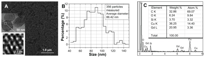
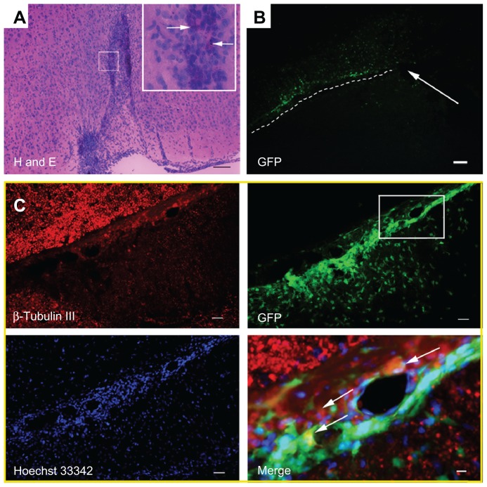
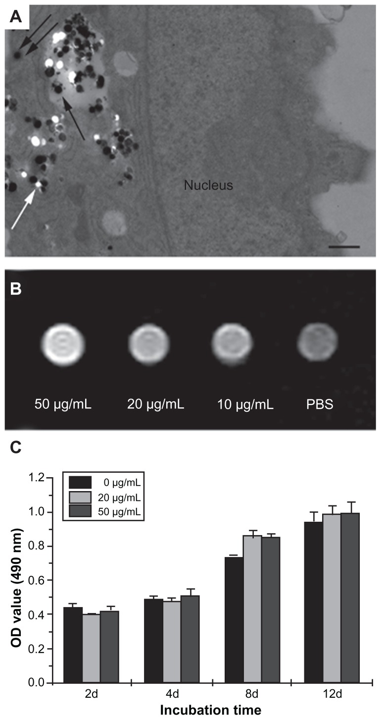
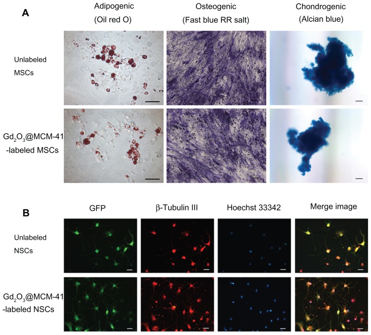
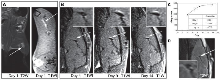
We investigated the tracking potential of a magnetic resonance imaging (MRI) probe made of gadolinium-doped mesoporous silica MCM-41 (Gd(2)O(3)@MCM-41) nanoparticles for transplanted bone mesenchymal stem cells (MSCs) and neural stem cells (NSCs) in vivo. The nanoparticles, synthesized using a one-step synthetic method, possess hexagonal mesoporous structures with appropriate assembly of nanoscale Gd(2)O(3) clusters. They show little cytotoxicity against proliferation and have a lower effect on the inherent differentiation potential of these labeled stem cells. The tracking of labeled NSCs in murine brains was dynamically determined with a clinical 3T MRI system for at least 14 days. The migration of labeled NSCs identified by MRI corresponded to the results of immunofluorescence imaging. Our study confirms that Gd(2)O(3)@MCM-41 particles can serve as an ideal vector for long-term MRI tracking of MSCs and NSCs in vivo.
我们研究了由掺杂钆的介孔硅 MCM-41(Gd2O3@MCM-41)纳米粒子制成的磁共振成像(MRI)探针在体内对移植的骨髓间充质干细胞(MSCs)和神经干细胞(NSCs)的跟踪潜力。这些纳米粒子使用一步合成法合成,具有适当组装的纳米级 Gd2O3 簇的六方介孔结构。它们对增殖的细胞毒性很小,对这些标记的干细胞的固有分化潜能的影响也较小。使用临床 3T MRI 系统至少 14 天来动态确定标记的 NSCs 在小鼠脑中的迁移情况。MRI 鉴定的标记 NSCs 的迁移与免疫荧光成像的结果一致。我们的研究证实,Gd2O3@MCM-41 颗粒可用作体内 MSC 和 NSCs 长期 MRI 跟踪的理想载体。