Hayashi Yasuhiko, Watanabe Takuya, Kita Daisuke, Hayashi Yutaka, Takahira Masayuki, Hamada Jun-Ichiro
Department of Neurosurgery, Graduate School of Medical Science, Kanazawa University, 13-1 Takara-machi, Kanazawa 920-8640, Japan.
Case Rep Ophthalmol Med. 2012;2012:604574. doi: 10.1155/2012/604574. Epub 2012 Dec 23.
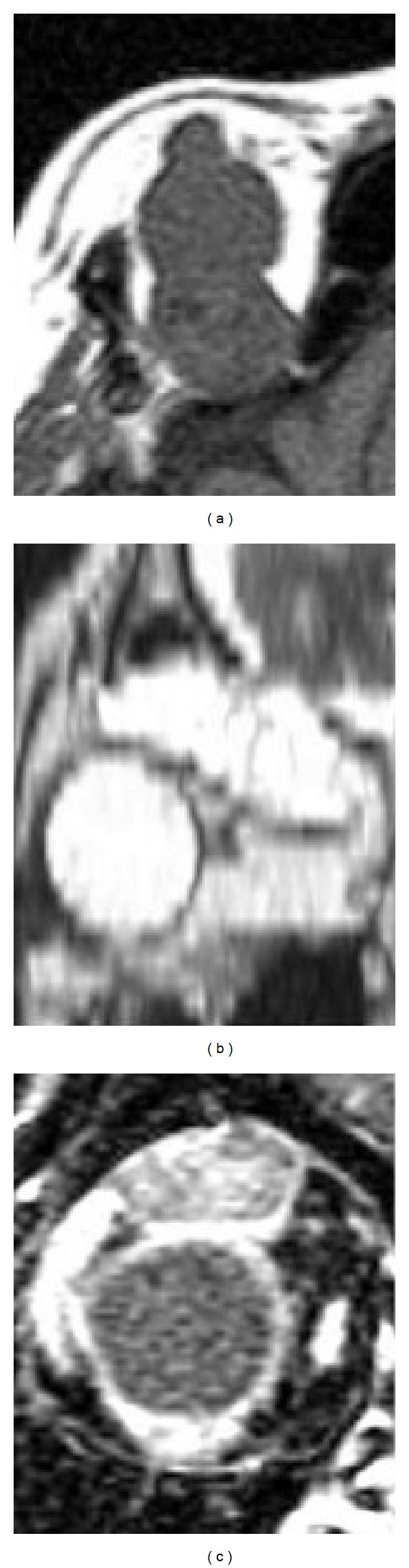
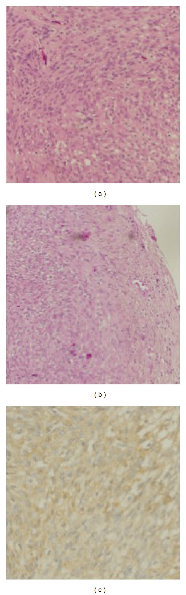
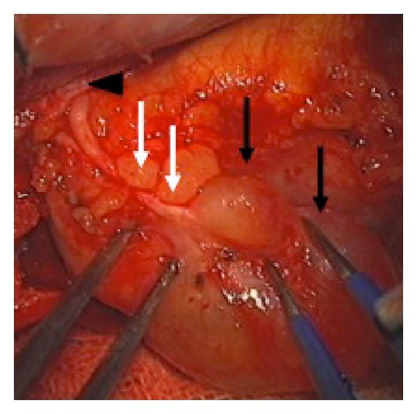
Schwannomas of the orbit are very rare benign neoplasms. Intraorbital cystic schwannomas originating from the frontal nerve are even rarer, with only 1 case reported to date. This is most likely due to the fact that, in most cases, the origin of the orbital schwannoma cannot be identified intraoperatively. The nerve origin is usually speculated from histological examination of the specimen and the postoperative neurological deficits of the patient. Here, we present the case of a 65-year-old woman with a one-month history of exophthalmos, whose orbital cystic lesion was completely removed by microsurgical transcranial operation. Intraoperatively, the continuity between the tumor and frontal nerve was seen macroscopically, leading us to confirm the frontal nerve as an origin of the tumor, which was consistent with the postoperative neurological findings. The diagnosis of the tumor was established as schwannoma from the histological examination. As a differential diagnosis of the orbital cystic lesions, the possibility of schwannomas should be kept in mind.
眼眶神经鞘瘤是非常罕见的良性肿瘤。起源于额神经的眶内囊性神经鞘瘤更为罕见,迄今为止仅报道过1例。这很可能是因为在大多数情况下,眼眶神经鞘瘤的起源在术中无法确定。神经起源通常是根据标本的组织学检查以及患者术后的神经功能缺损来推测的。在此,我们报告1例65岁女性患者,有1个月的眼球突出病史,其眼眶囊性病变通过显微经颅手术被完全切除。术中肉眼可见肿瘤与额神经之间的连续性,这使我们确认额神经为肿瘤的起源,这与术后神经学检查结果相符。经组织学检查确诊该肿瘤为神经鞘瘤。作为眼眶囊性病变的鉴别诊断,应考虑神经鞘瘤的可能性。