• 文献检索
  • 文档翻译
  • 深度研究
  • 学术资讯
  • Suppr Zotero 插件Zotero 插件
  • 邀请有礼
  • 套餐&价格
  • 历史记录
应用&插件
Suppr Zotero 插件Zotero 插件浏览器插件Mac 客户端Windows 客户端微信小程序
定价
高级版会员购买积分包购买API积分包
服务
文献检索文档翻译深度研究API 文档MCP 服务
关于我们
关于 Suppr公司介绍联系我们用户协议隐私条款
关注我们

Suppr 超能文献

核心技术专利:CN118964589B侵权必究
粤ICP备2023148730 号-1Suppr @ 2026

文献检索

告别复杂PubMed语法,用中文像聊天一样搜索,搜遍4000万医学文献。AI智能推荐,让科研检索更轻松。

立即免费搜索

文件翻译

保留排版,准确专业,支持PDF/Word/PPT等文件格式,支持 12+语言互译。

免费翻译文档

深度研究

AI帮你快速写综述,25分钟生成高质量综述,智能提取关键信息,辅助科研写作。

立即免费体验

意大利正畸患者上颌窦偶然发现的患病率:一项回顾性锥形束计算机断层扫描研究

Prevalence of incidental maxillary sinus findings in Italian orthodontic patients: a retrospective cone-beam computed tomography study.

作者信息

Gracco Antonio, Incerti Parenti Serena, Ioele Christian, Alessandri Bonetti Giulio, Stellini Edoardo

机构信息

Department of Orthodontics, School of Dentistry, University of Padua, Padua, Italy.

出版信息

Korean J Orthod. 2012 Dec;42(6):329-34. doi: 10.4041/kjod.2012.42.6.329. Epub 2012 Dec 28.

DOI:10.4041/kjod.2012.42.6.329
PMID:23323247
原文链接:https://pmc.ncbi.nlm.nih.gov/articles/PMC3542453/
Abstract

OBJECTIVES

To determine the prevalence of incidental maxillary sinus findings in a large sample of orthodontic patients by cone-beam computed tomography (CBCT) with a wide field of view and assess the relationships of such abnormalities with age and gender.

METHODS

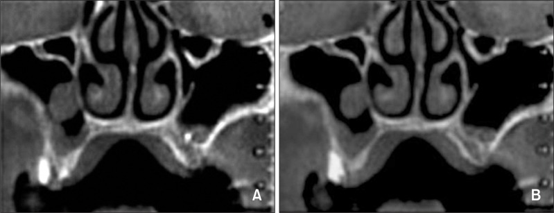
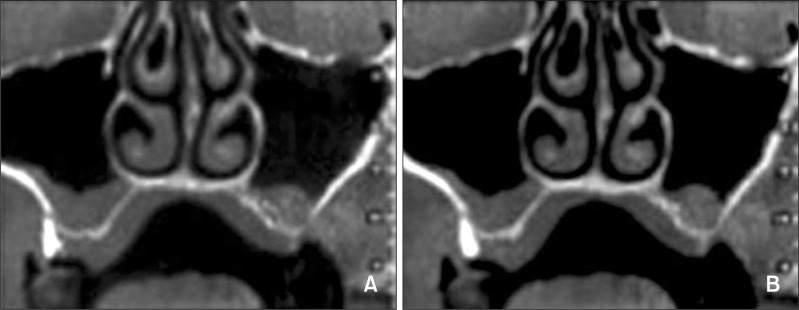
Five hundred thirteen CBCT scans obtained for orthodontic diagnosis and treatment planning in a Northern Italian population (N = 513; 292 female and 221 male subjects; 1,026 maxillary sinuses) were studied. The frequencies of pseudocysts and mucosal thickening of the maxillary sinus were recorded. Logistic regression analysis was used to determine the influence of age and gender on these abnormalities.

RESULTS

Pseudocysts were detected in 52 patients (10.1%) and 59 sinuses (5.75%). Mucosal thickening was observed in 206 patients (40.1%) and 258 sinuses (25.1%). Gender and age were significantly associated with pseudocysts (p = 0.027) and mucosal thickening (p < 0.001), respectively.

CONCLUSIONS

Half of the orthodontic patients had incidental maxillary sinus findings. Men were more likely to show pseudocysts, and older patients (aged 41 - 60 years) were more likely to show mucosal thickening.

摘要

目的

通过大视野锥形束计算机断层扫描(CBCT)确定大量正畸患者中上颌窦偶然发现的患病率,并评估这些异常与年龄和性别的关系。

方法

研究了在意大利北部人群中为正畸诊断和治疗计划而获取的513例CBCT扫描(N = 513;292名女性和221名男性受试者;1026个上颌窦)。记录上颌窦假性囊肿和黏膜增厚的频率。采用逻辑回归分析来确定年龄和性别对这些异常的影响。

结果

在52例患者(10.1%)和59个鼻窦(5.75%)中检测到假性囊肿。在206例患者(40.1%)和258个鼻窦(25.1%)中观察到黏膜增厚。性别和年龄分别与假性囊肿(p = 0.027)和黏膜增厚(p < 0.001)显著相关。

结论

一半的正畸患者有上颌窦偶然发现。男性更易出现假性囊肿,年龄较大的患者(41 - 60岁)更易出现黏膜增厚。

https://cdn.ncbi.nlm.nih.gov/pmc/blobs/cb30/3542453/e2dc1f9f83c2/kjod-42-329-g002.jpg
https://cdn.ncbi.nlm.nih.gov/pmc/blobs/cb30/3542453/b079d18cf0c5/kjod-42-329-g001.jpg
https://cdn.ncbi.nlm.nih.gov/pmc/blobs/cb30/3542453/e2dc1f9f83c2/kjod-42-329-g002.jpg
https://cdn.ncbi.nlm.nih.gov/pmc/blobs/cb30/3542453/b079d18cf0c5/kjod-42-329-g001.jpg
https://cdn.ncbi.nlm.nih.gov/pmc/blobs/cb30/3542453/e2dc1f9f83c2/kjod-42-329-g002.jpg

相似文献

1
Prevalence of incidental maxillary sinus findings in Italian orthodontic patients: a retrospective cone-beam computed tomography study.意大利正畸患者上颌窦偶然发现的患病率:一项回顾性锥形束计算机断层扫描研究
Korean J Orthod. 2012 Dec;42(6):329-34. doi: 10.4041/kjod.2012.42.6.329. Epub 2012 Dec 28.
2
Incidental maxillary sinus findings in orthodontic patients: a radiographic analysis using cone-beam computed tomography (CBCT).正畸患者上颌窦偶然发现:锥形束 CT(CBCT)的放射分析。
Orthod Craniofac Res. 2011 Feb;14(1):17-24. doi: 10.1111/j.1601-6343.2010.01502.x. Epub 2010 Nov 22.
3
Prevalence of Incidental Findings and Assessment of Maxillary Sinus Pathologies and Dental Diseases Using Cone-Beam Computed Tomography (CBCT) in the Tamil Nadu Population: A Retrospective Study.泰米尔纳德邦人群中使用锥形束计算机断层扫描(CBCT)对上颌窦病变和牙科疾病的偶然发现患病率及评估:一项回顾性研究
Cureus. 2024 Sep 8;16(9):e68929. doi: 10.7759/cureus.68929. eCollection 2024 Sep.
4
Prevalence of incidental maxillary sinus pathologies in dental patients on cone-beam computed tomographic images.牙科患者锥形束计算机断层扫描图像上上颌窦偶发病变的患病率。
Contemp Clin Dent. 2014 Jul;5(3):361-5. doi: 10.4103/0976-237X.137949.
5
Cone-beam computed tomographic evidence of the association between periodontal bone loss and mucosal thickening of the maxillary sinus.锥形束计算机断层扫描证据表明牙周骨丧失与上颌窦黏膜增厚之间存在关联。
J Periodontol. 2012 May;83(5):557-64. doi: 10.1902/jop.2011.110376. Epub 2011 Sep 12.
6
Prevalence of pathologic findings in the floor of the maxillary sinuses on cone beam computed tomography images.锥形束计算机断层扫描图像上上颌窦底病理表现的患病率。
Gen Dent. 2017 Mar-Apr;65(2):28-32.
7
Association between odontogenic conditions and maxillary sinus mucosal thickening: a retrospective CBCT study.牙源性疾病与上颌窦黏膜增厚的相关性:一项回顾性 CBCT 研究。
Clin Oral Investig. 2019 Jan;23(1):123-131. doi: 10.1007/s00784-018-2418-x. Epub 2018 Mar 17.
8
Associations between maxillary sinus mucosal thickening and apical periodontitis using cone-beam computed tomography scanning: a retrospective study.应用锥形束 CT 扫描研究上颌窦黏膜增厚与根尖周炎的相关性:一项回顾性研究。
J Endod. 2012 Aug;38(8):1069-74. doi: 10.1016/j.joen.2012.04.027. Epub 2012 Jun 20.
9
Frequency and type of incidentally detected radiodensities in the maxillary sinus: a retrospective analysis using cone beam computed tomography (CBCT).上颌窦偶然发现的放射性密度的频率和类型:使用锥形束计算机断层扫描(CBCT)的回顾性分析。
Clin Oral Investig. 2019 Mar;23(3):1091-1099. doi: 10.1007/s00784-018-2541-8. Epub 2018 Jun 28.
10
Incidence of Maxillary Sinus Disorders in Dental Patients Undergoing Cone-Beam Computed Tomography: A Retrospective Cross-Sectional Study.接受锥形束计算机断层扫描的牙科患者上颌窦疾病的发病率:一项回顾性横断面研究。
Cureus. 2024 Oct 8;16(10):e71114. doi: 10.7759/cureus.71114. eCollection 2024 Oct.

引用本文的文献

1
Incidence of Maxillary Sinus Disorders in Dental Patients Undergoing Cone-Beam Computed Tomography: A Retrospective Cross-Sectional Study.接受锥形束计算机断层扫描的牙科患者上颌窦疾病的发病率:一项回顾性横断面研究。
Cureus. 2024 Oct 8;16(10):e71114. doi: 10.7759/cureus.71114. eCollection 2024 Oct.
2
Prevalence of Incidental Findings and Assessment of Maxillary Sinus Pathologies and Dental Diseases Using Cone-Beam Computed Tomography (CBCT) in the Tamil Nadu Population: A Retrospective Study.泰米尔纳德邦人群中使用锥形束计算机断层扫描(CBCT)对上颌窦病变和牙科疾病的偶然发现患病率及评估:一项回顾性研究
Cureus. 2024 Sep 8;16(9):e68929. doi: 10.7759/cureus.68929. eCollection 2024 Sep.
3

本文引用的文献

1
Cone-beam computed tomographic evidence of the association between periodontal bone loss and mucosal thickening of the maxillary sinus.锥形束计算机断层扫描证据表明牙周骨丧失与上颌窦黏膜增厚之间存在关联。
J Periodontol. 2012 May;83(5):557-64. doi: 10.1902/jop.2011.110376. Epub 2011 Sep 12.
2
Clinical effectiveness of orthodontic miniscrew implants: a meta-analysis.正畸微螺钉种植体的临床效果:荟萃分析。
J Dent Res. 2011 Aug;90(8):969-76. doi: 10.1177/0022034511409236. Epub 2011 May 18.
3
Prevalence of pathologic findings in the maxillary sinus in cone-beam computerized tomography.
Retrospective analysis of pathological changes in the maxillary sinus with CBCT.
CBCT 上颌窦病变的回顾性分析。
Sci Rep. 2024 Jul 5;14(1):15529. doi: 10.1038/s41598-024-66527-7.
4
Prevalence and Clinical Significance of Incidental Findings in the Maxillofacial Complex of Adolescent Orthodontic Patients: A Retrospective Cone Beam Computed Tomography Analysis.青少年正畸患者颌面复合体中偶然发现的患病率及临床意义:一项回顾性锥形束计算机断层扫描分析
Cureus. 2023 Oct 22;15(10):e47480. doi: 10.7759/cureus.47480. eCollection 2023 Oct.
5
Comparison of Diagnostic Accuracy of Panoramic Radiography to CBCT in Maxillary Sinus Diseases.全景X线摄影与锥形束CT在上颌窦疾病诊断准确性的比较
Indian J Otolaryngol Head Neck Surg. 2023 Apr;75(Suppl 1):881-892. doi: 10.1007/s12070-023-03475-0. Epub 2023 Jan 18.
6
The Relationship between an Accessory Maxillary Ostium and Variations in Structures Adjacent to the Maxillary Sinus without Polyps.副上颌窦口与无息肉上颌窦相邻结构变异之间的关系
Int Arch Otorhinolaryngol. 2022 Jan 28;26(4):e548-e555. doi: 10.1055/s-0042-1742325. eCollection 2022 Oct.
7
Prevalence of incidental findings in adult vs. adolescent patients in the course of orthodontic X-ray diagnostics.正畸 X 射线诊断中成年患者与青少年患者偶然发现的发生率比较。
J Orofac Orthop. 2023 Sep;84(5):298-310. doi: 10.1007/s00056-022-00399-2. Epub 2022 May 18.
8
Odontogenic Maxillary Sinusopathies: a Radiological Classification.牙源性上颌窦病变:一种放射学分类
J Maxillofac Oral Surg. 2022 Mar;21(1):141-149. doi: 10.1007/s12663-020-01329-8. Epub 2020 Jan 16.
9
Effect of Schneiderian Membrane Thickening on the Maxillary Sinus Augmentation and Implantation Outcomes: A Systematic Review.筛窦黏膜增厚对上颌窦提升及种植效果的影响:一项系统评价
J Maxillofac Oral Surg. 2021 Dec;20(4):534-544. doi: 10.1007/s12663-021-01551-y. Epub 2021 Apr 2.
10
Incidental findings on cone-beam computed tomographic images: paranasal sinus findings and nasal septum variations.锥形束计算机断层扫描图像上的偶然发现:鼻窦发现及鼻中隔变异
Oral Radiol. 2018 Jan;34(1):40-48. doi: 10.1007/s11282-017-0283-y. Epub 2017 Mar 27.
锥形束计算机断层扫描在上颌窦中的病理发现患病率
Oral Surg Oral Med Oral Pathol Oral Radiol Endod. 2011 May;111(5):634-40. doi: 10.1016/j.tripleo.2010.12.007. Epub 2011 Mar 27.
4
Incidental findings arising with cone beam computed tomography imaging of the orthodontic patient.正畸患者锥形束 CT 成像中的偶然发现。
Angle Orthod. 2011 Mar;81(2):350-5. doi: 10.2319/032210-165.1.
5
Incidental maxillary sinus findings in orthodontic patients: a radiographic analysis using cone-beam computed tomography (CBCT).正畸患者上颌窦偶然发现:锥形束 CT(CBCT)的放射分析。
Orthod Craniofac Res. 2011 Feb;14(1):17-24. doi: 10.1111/j.1601-6343.2010.01502.x. Epub 2010 Nov 22.
6
Miniscrew insertion and the maxillary sinus: an endoscopic evaluation.微型螺钉植入与上颌窦:一项内镜评估
J Clin Orthod. 2010 Jul;44(7):439-43.
7
Prevalence of mucosal abnormalities of the maxillary sinus and their relationship to dental disease in panoramic radiography: results from the Health 2000 Health Examination Survey.全景X线片中上颌窦黏膜异常的患病率及其与牙病的关系:来自2000年健康检查调查的结果
Oral Surg Oral Med Oral Pathol Oral Radiol Endod. 2010 Mar;109(3):e80-7. doi: 10.1016/j.tripleo.2009.10.031.
8
The incidence of maxillary sinus retention cysts in orthodontic patients.正畸患者上颌窦潴留囊肿的发病率。
World J Orthod. 2009 Summer;10(2):e7-8.
9
Basic principles for use of dental cone beam computed tomography: consensus guidelines of the European Academy of Dental and Maxillofacial Radiology.牙科锥形束计算机断层扫描使用的基本原则:欧洲口腔颌面放射学会共识指南
Dentomaxillofac Radiol. 2009 May;38(4):187-95. doi: 10.1259/dmfr/74941012.
10
American Academy of Oral and Maxillofacial Radiology executive opinion statement on performing and interpreting diagnostic cone beam computed tomography.美国口腔颌面放射学会关于进行和解读诊断性锥形束计算机断层扫描的执行意见声明
Oral Surg Oral Med Oral Pathol Oral Radiol Endod. 2008 Oct;106(4):561-2. doi: 10.1016/j.tripleo.2008.07.007.