Institute of Genetic Medicine, Newcastle University, International Center for Life, Newcastle, UK.
J Cardiovasc Magn Reson. 2013 Jan 16;15(1):4. doi: 10.1186/1532-429X-15-4.
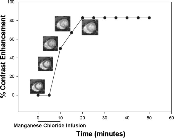
Manganese-enhanced cardiovascular magnetic resonance (MECMR) can non-invasively assess myocardial calcium influx, and calcium levels are known to be elevated in muscular dystrophy cardiomyopathy based on cellular studies.
Left ventricular functional studies and MECMR were performed in mdx mice (model of Duchenne muscular dystrophy, 24 and 40 weeks) and Sgcd -/- mice (limb girdle muscular dystrophy 2 F, 16 and 32 weeks), compared to wild type controls (C57Bl/10, WT).
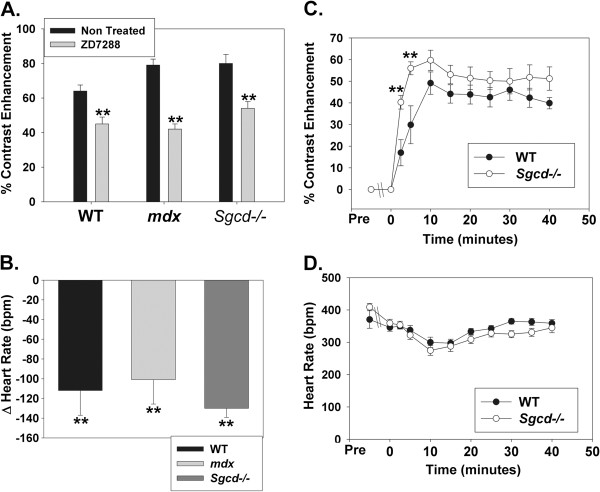
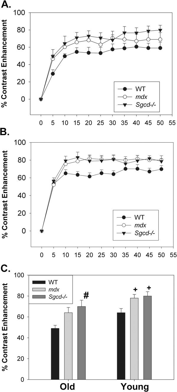
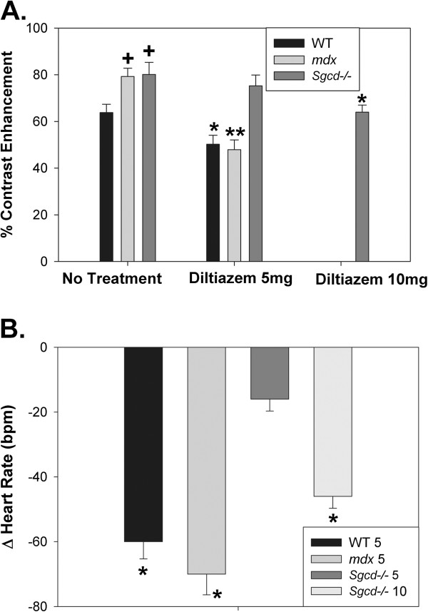
Both models had left ventricular hypertrophy at the later age compared to WT, though the mdx mice had reduced stroke volumes and the Sgcd -/- mice increased heart rate and cardiac index. Especially at the younger ages, MECMR was significantly elevated in both models (both P < 0.05 versus WT). The L-type calcium channel inhibitor diltiazem (5 mg/kg i.p.) significantly reduced MECMR in the mdx mice (P < 0.01), though only with a higher dose (10 mg/kg i.p.) in the Sgcd -/- mice (P < 0.05). As the Sgcd -/- mice had increased heart rates, to determine the role of heart rate in MECMR we studied the hyperpolarization-activated cyclic nucleotide-gated channel inhibitor ZD 7288 which selectively reduces heart rate. This reduced heart rate and MECMR in all mouse groups. However, when looking at the time course of reduction of MECMR in the Sgcd -/- mice at up to 5 minutes of the manganese infusion when heart rates were matched to the WT mice, MECMR was still significantly elevated in the Sgcd -/- mice (P < 0.01) indicating that heart rate alone could not account for all the increased MECMR.
Despite both mouse models exhibiting increased in-vivo calcium influx at an early stage in the development of the cardiomyopathy before left ventricular hypertrophy, there are distinct phenotypical differences between the 2 models in terms of heart rates, hemodynamics and responses to calcium channel inhibitors.
锰增强心血管磁共振(MECMR)可无创评估心肌钙内流,基于细胞研究已知钙水平在肌肉营养不良性心肌病中升高。
在 mdx 小鼠(杜氏肌营养不良症模型,24 周和 40 周)和 Sgcd -/- 小鼠(肢带型肌营养不良症 2F,16 周和 32 周)以及野生型对照(C57Bl/10,WT)中进行左心室功能研究和 MECMR。
与 WT 相比,两种模型在后期都有左心室肥厚,但 mdx 小鼠的每搏量减少,而 Sgcd -/- 小鼠的心率增加和心脏指数增加。特别是在较年轻的年龄,两种模型的 MECMR 均显著升高(均 P < 0.05 与 WT 相比)。L 型钙通道抑制剂地尔硫卓(5mg/kg ip)可显著降低 mdx 小鼠的 MECMR(P < 0.01),但在 Sgcd -/- 小鼠中需要更高剂量(10mg/kg ip)(P < 0.05)。由于 Sgcd -/- 小鼠的心率增加,为了确定心率在 MECMR 中的作用,我们研究了选择性降低心率的超极化激活环核苷酸门控通道抑制剂 ZD 7288。这降低了所有小鼠组的心率和 MECMR。然而,当观察到 Sgcd -/- 小鼠中锰输注长达 5 分钟时 MECMR 降低的时间过程时,当心率与 WT 小鼠匹配时,Sgcd -/- 小鼠中的 MECMR 仍然显著升高(P < 0.01),表明心率单独不能解释所有增加的 MECMR。
尽管两种小鼠模型在心肌病发展的早期阶段就表现出钙内流增加,但在心率、血液动力学和钙通道抑制剂反应方面,两种模型之间存在明显的表型差异。