Experimental Molecular Imaging, Department of Radiology, Leiden University Medical Center, Leiden, The Netherlands.
Cell Death Dis. 2013 Jan 24;4(1):e473. doi: 10.1038/cddis.2012.207.
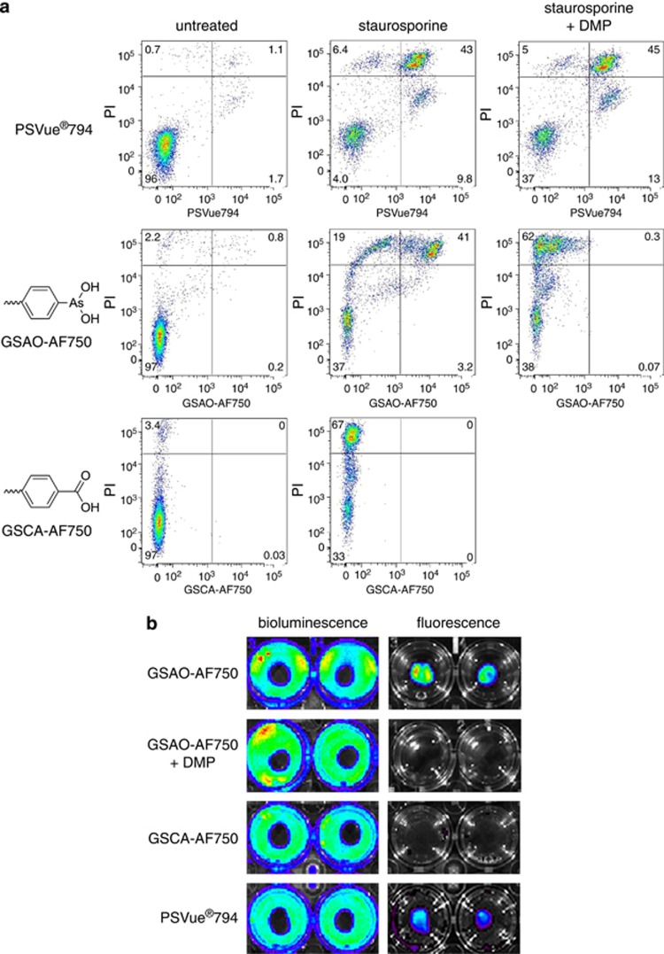
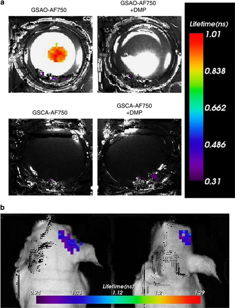
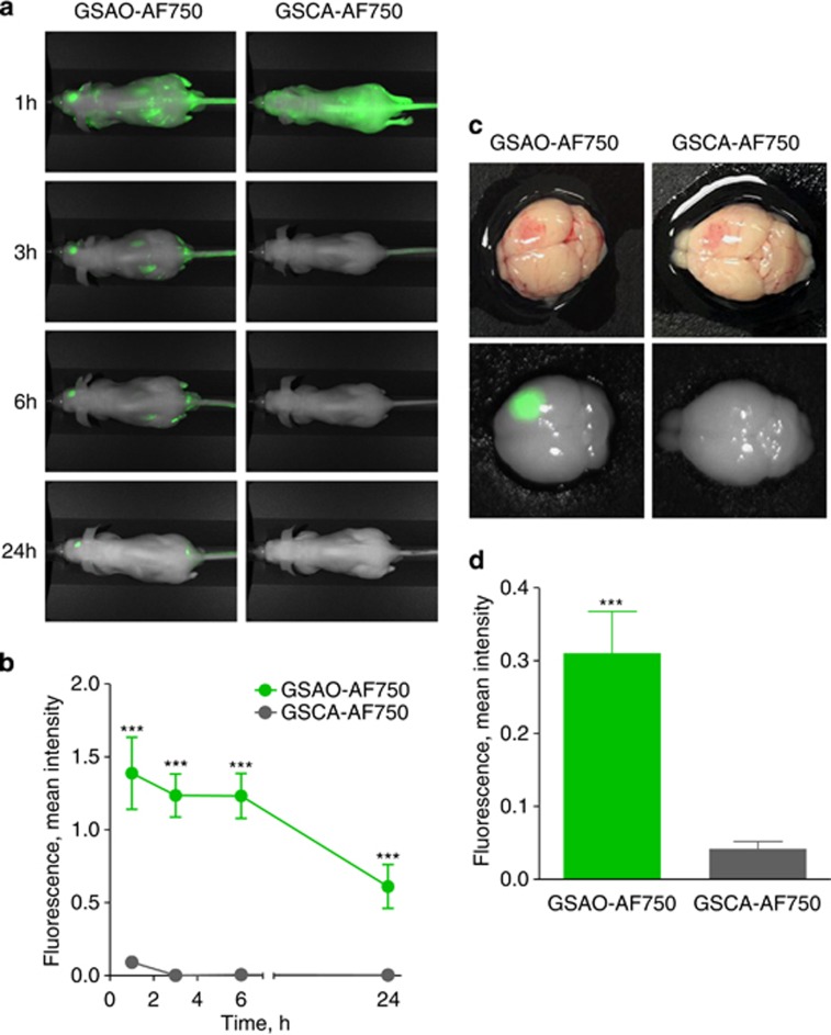
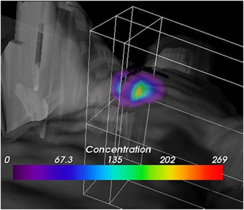
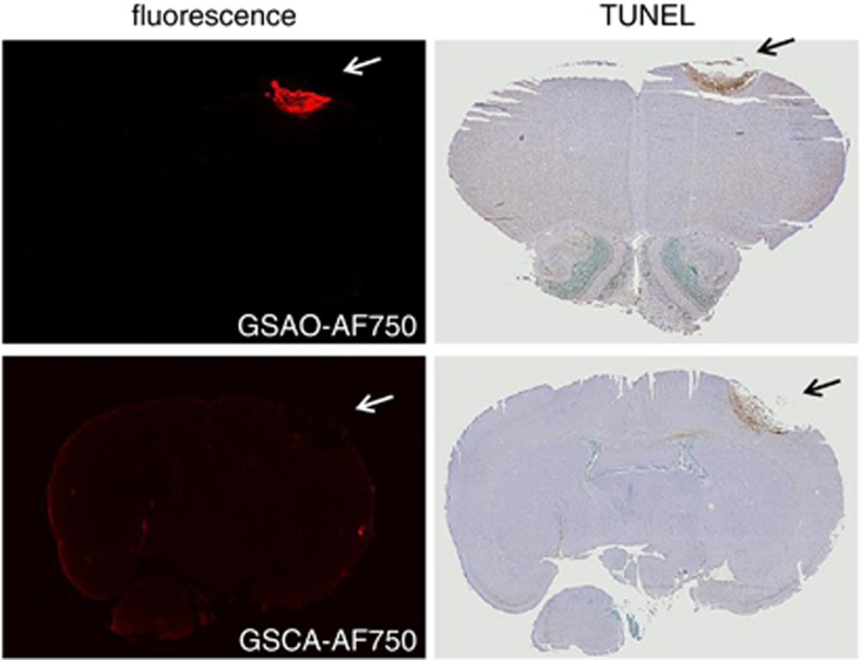
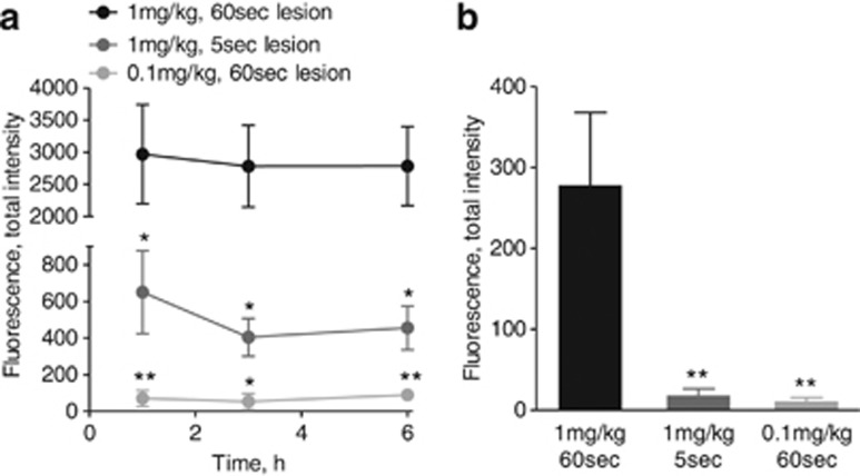
Traumatic brain injury is a major public health concern and is characterised by both apoptotic and necrotic cell death in the lesion. Anatomical imaging is usually used to assess traumatic brain injuries and there is a need for imaging modalities that provide complementary cellular information. We sought to non-invasively image cell death in a mouse model of traumatic brain injury using a near-infrared fluorescent conjugate of a synthetic heat shock protein-90 alkylator, 4-(N-(S-glutathionylacetyl) amino) phenylarsonous acid (GSAO). GSAO labels both apoptotic and necrotic cells coincident with loss of plasma membrane integrity. The optical GSAO specifically labelled apoptotic and necrotic cells in culture and did not accumulate in healthy organs or tissues in the living mouse body. The conjugate is a very effective imager of cell death in brain lesions. The optical GSAO was detected by fluorescence intensity and GSAO bound to dying/dead cells was detected from prolongation of the fluorescence lifetime. An optimal signal-to-background ratio was achieved as early as 3 h after injection of the probe and the signal intensity positively correlated with both lesion size and probe concentration. This optical GSAO offers a convenient and robust means to non-invasively image apoptotic and necrotic cell death in brain and other lesions.
创伤性脑损伤是一个主要的公共卫生关注点,其特征在于病变部位同时存在细胞凋亡和坏死。解剖成像通常用于评估创伤性脑损伤,需要成像方式来提供互补的细胞信息。我们试图使用合成热休克蛋白 90 烷化剂 4-(N-(S-谷胱甘肽乙酰基)氨基)苯砷酸(GSAO)的近红外荧光缀合物,非侵入性地对创伤性脑损伤的小鼠模型中的细胞死亡进行成像。GSAO 标记同时伴有质膜完整性丧失的凋亡和坏死细胞。光学 GSAO 可特异性标记培养物中的凋亡和坏死细胞,并且不会在活体小鼠的健康器官或组织中积聚。该缀合物是脑损伤中细胞死亡的非常有效的成像剂。通过荧光强度检测光学 GSAO,通过荧光寿命延长检测与死亡/坏死细胞结合的 GSAO。在注射探针后 3 小时即可获得最佳的信噪比,并且信号强度与病变大小和探针浓度呈正相关。这种光学 GSAO 为非侵入性地对脑和其他病变中的凋亡和坏死细胞死亡进行成像提供了一种方便且强大的手段。