Schools of Dentistry, and Medical Science, Griffith Health Institute, Griffith University, Parklands Drive, Southport, Gold Coast, QLD, 4222, Australia.
Clin Exp Metastasis. 2013 Jun;30(5):659-70. doi: 10.1007/s10585-013-9570-0. Epub 2013 Feb 2.
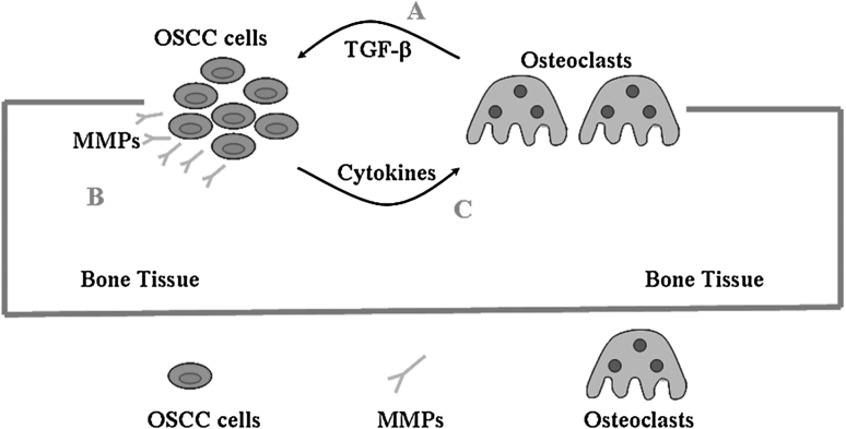
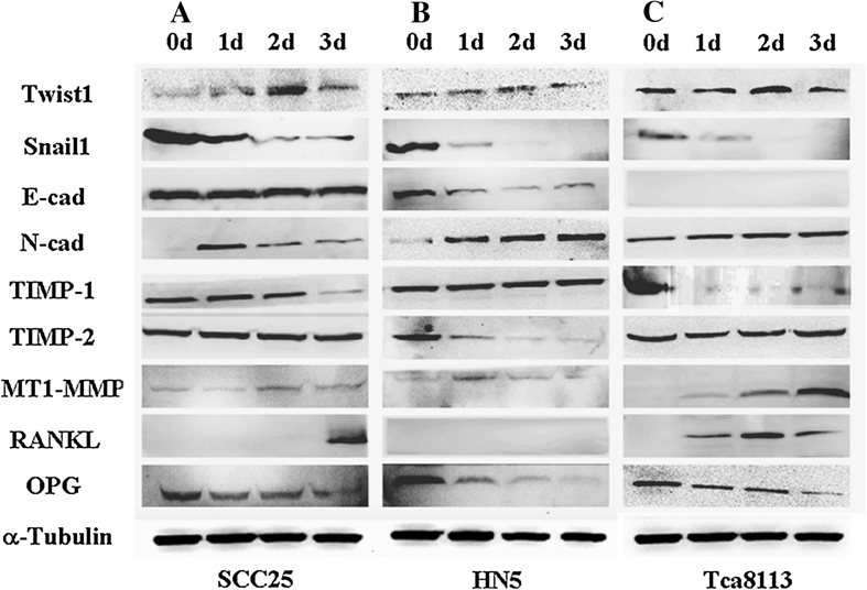
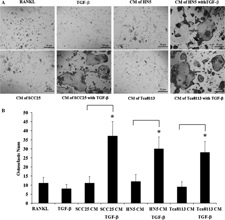
This study investigates relationships between EMT and bone invasion by OSCC. Three OSCC cell lines, SCC25, HN5, and Tca8113 were artificially induced to display EMT by adding 5 ng/mL of TGF-β1 to culture media for 1-3 days. Cell morphology and phenotypic changes was examined by immunocytochemical staining of CK and VIM. EMT markers, cell-invasion factors, and osteoclast-related molecules were analysed at mRNA, gelatine and protein levels by real-time PCR, gelatine zymography and Western blotting respectively. Mature osteoclasts differentiated from Raw264.7 cells were treated by conditioned medium (CM) of OSCC cells with/without TGF-β1. Immunohistochemistry was performed to validate proteins of CK, VIM, E-cad and Snail1 in OSCC tissue samples with bone invasion. Results showed minimal staining of VIM was found in SCC25 and HN5, while Tca8113 cells stained strongly. EMT markers Twist1 and N-cad were up-regulated; Snail1 and E-cad down-regulated in all cells. Of factors associated with invasion, MMP-2 was unchanged and MMP-9 increased in SCC25 and Tca8113, while MMP-2 was increased and MMP-9 unchanged in HN5. For osteoclast-related molecules, both MT1-MMP and RANKL were up-regulated, while OPG was down-regulated in all cells. CM of OSCC cells pre-treated with TGF-β1 showed to prolong survival of osteoclasts up to 4 days. All target molecules were validated in OSCC samples of bone invasion. These findings suggest that TGF-β1 not only induces EMT to increase the capacity of OSCC for invasion, but also promotes factors which prolong osteoclast survival. TGF-β1 may enhance the ability of MMP2/9 in resorbing bone and favouring invasion of cancer cells.
本研究调查 EMT 与口腔鳞状细胞癌(OSCC)骨侵袭之间的关系。通过在培养基中添加 5ng/mL TGF-β1 1-3 天,将三种 OSCC 细胞系 SCC25、HN5 和 Tca8113 人为诱导 EMT。通过 CK 和 VIM 的免疫细胞化学染色检查细胞形态和表型变化。通过实时 PCR、凝胶酶谱和 Western 印迹分别在 mRNA、凝胶酶和蛋白质水平上分析 EMT 标志物、细胞侵袭因子和破骨细胞相关分子。用 TGF-β1 处理的 OSCC 细胞条件培养基(CM)处理来自 Raw264.7 细胞的成熟破骨细胞。用免疫组织化学法验证具有骨侵袭的 OSCC 组织样本中 CK、VIM、E-cad 和 Snail1 蛋白。结果显示 SCC25 和 HN5 中 VIM 染色极少,而 Tca8113 细胞染色强烈。EMT 标志物 Twist1 和 N-cad 上调;所有细胞中 Snail1 和 E-cad 下调。与侵袭相关的因子中,MMP-2 不变,MMP-9 在 SCC25 和 Tca8113 中增加,而 MMP-2 增加,MMP-9 在 HN5 中不变。对于破骨细胞相关分子,MT1-MMP 和 RANKL 均上调,而所有细胞中 OPG 下调。经 TGF-β1 预处理的 OSCC 细胞 CM 显示可将破骨细胞的存活时间延长至 4 天。在具有骨侵袭的 OSCC 样本中验证了所有靶分子。这些发现表明,TGF-β1 不仅诱导 EMT 增加 OSCC 的侵袭能力,还促进延长破骨细胞存活的因子。TGF-β1 可能增强 MMP2/9 分解骨的能力,并有利于癌细胞侵袭。