Guanghua School of Stomatology, Hospital of Stomatology, Sun Yat-sen University and Guangdong Provincial Key Laboratory of Stomatology, Guangzhou, Guangdong 510080, P.R. China.
Affiliated High School-South China Normal University, Guangzhou, Guangdong 510630, P.R. China.
Oncol Rep. 2018 Mar;39(3):1043-1051. doi: 10.3892/or.2017.6166. Epub 2017 Dec 19.
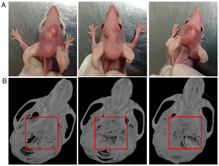
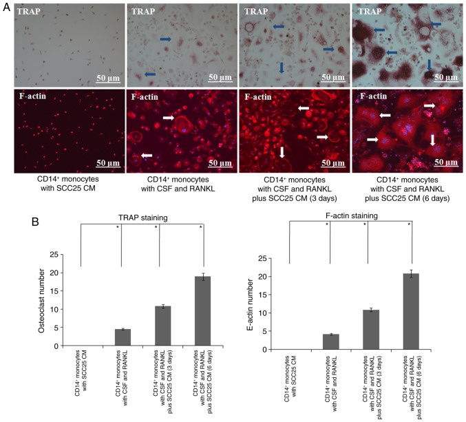
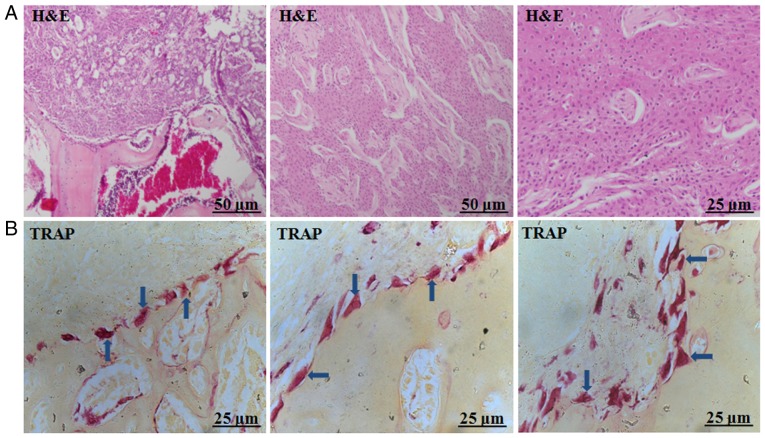
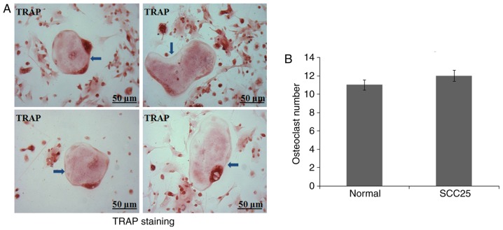
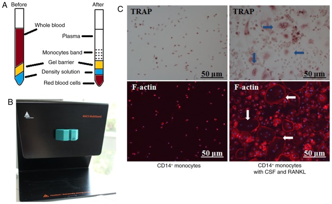
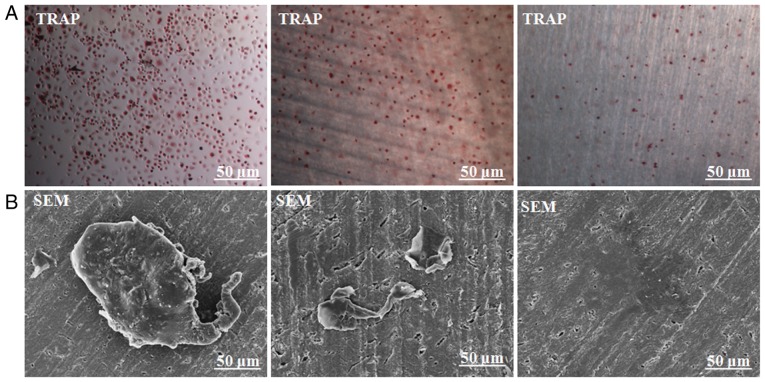
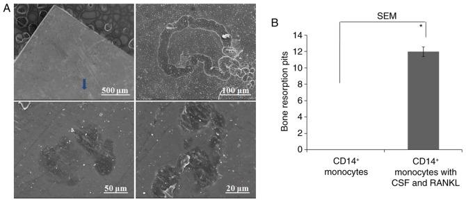
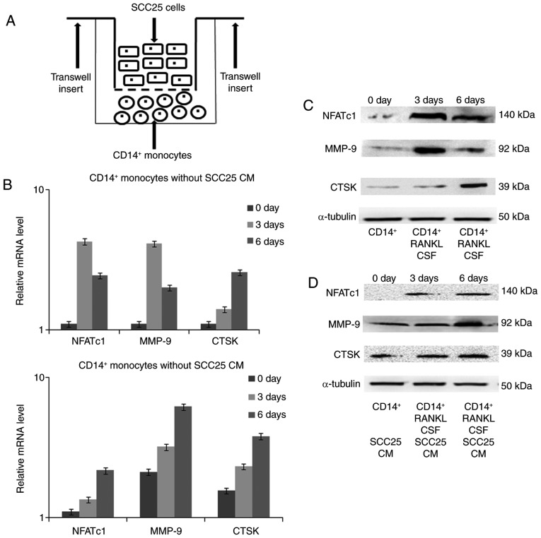
The present study aimed to characterize different phenotypes of osteoclasts in the progression of bone invasion by oral squamous cell carcinoma (OSCC). A local bone invasion model of OSCC was established by injecting SCC25 human OSCC cells into the center of calvariae in nude mice, and all mice were found to have a typical bone resorption area. Staining for tartrate-resistant acid phosphatase (TRAP) revealed various types of giant osteoclasts in the tumour-bone interface. Bone marrow cells (BMCs) were isolated from the nude mice for primary osteoclast culture, but only a few giant osteoclasts were generated. Additionally, special blood centrifuge tubes were utilized to obtain large numbers of peripheral blood mononuclear cells (PBMCs). Using magnetic activated cell sorting (MACS) and the cytokines colony-stimulating factor (CSF) and receptor activator of nuclear factor-κb ligand (RANKL), we differentiated human osteoclasts from CD14+ monocytes of PBMCs. Bone resorption was further confirmed by a bone resorption assay. Finally, Transwell inserts were used for indirect cell co-culture of SCC25 cells and CD14+ monocytes. Expression of specific osteoclast markers was detected by real-time PCR and western blotting. After co-culture for 3 and 6 days, conditioned medium (CM) of SCC25 cells stimulated the expression of osteoclast markers, and additional osteoclasts were detected through staining of TRAP and F-actin. In the present study distinct osteoclast phenotypes were observed in the established bone invasion animal model, and were confirmed using various primary osteoclast cultures. CM of OSCC cells may promote the expression of osteoclast markers and induce the differentiation of monocytes to mature osteoclasts, which can resorb adjacent bone tissue.
本研究旨在探讨口腔鳞状细胞癌(OSCC)侵袭骨过程中破骨细胞的不同表型特征。通过将 SCC25 人 OSCC 细胞注入裸鼠颅骨中心,建立了局部骨侵袭 OSCC 模型,所有小鼠均出现典型的骨吸收区。抗酒石酸酸性磷酸酶(TRAP)染色显示肿瘤-骨界面存在各种类型的巨大破骨细胞。从裸鼠骨髓中分离骨髓细胞(BMCs)进行原代破骨细胞培养,但仅产生少数巨大破骨细胞。此外,还使用特殊的血液离心管从外周血单核细胞(PBMCs)中获得大量的外周血单核细胞。利用磁激活细胞分选(MACS)和细胞因子集落刺激因子(CSF)和核因子-κb 配体受体激活剂(RANKL),我们从 PBMCs 的 CD14+单核细胞中分化出人类破骨细胞。通过骨吸收测定进一步证实了骨吸收。最后,使用 Transwell 插入物进行 SCC25 细胞和 CD14+单核细胞的间接细胞共培养。通过实时 PCR 和 Western blot 检测破骨细胞特异性标志物的表达。共培养 3 天和 6 天后,SCC25 细胞的条件培养基(CM)刺激破骨细胞标志物的表达,并通过 TRAP 和 F-actin 染色检测到额外的破骨细胞。在本研究中,在建立的骨侵袭动物模型中观察到了不同的破骨细胞表型,并通过各种原代破骨细胞培养得到了证实。OSCC 细胞的 CM 可能促进破骨细胞标志物的表达,并诱导单核细胞向成熟破骨细胞分化,从而吸收周围的骨组织。