Molecular Imaging and Translational Research, University of Tennessee Medical Center, Knoxville, TN 37920, USA.
Phys Med Biol. 2013 Mar 7;58(5):1465-78. doi: 10.1088/0031-9155/58/5/1465. Epub 2013 Feb 13.
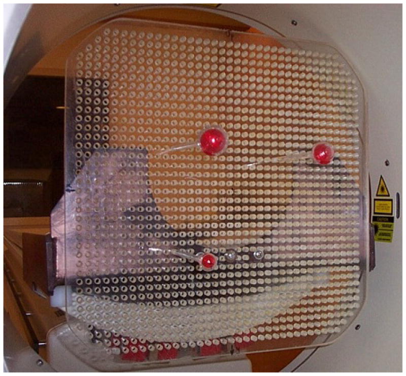
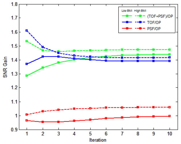
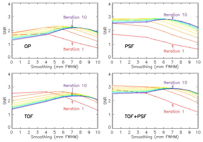
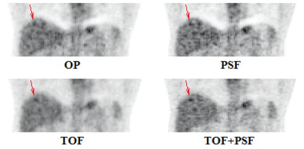
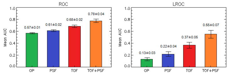
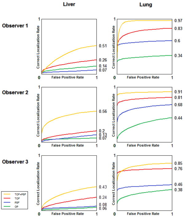
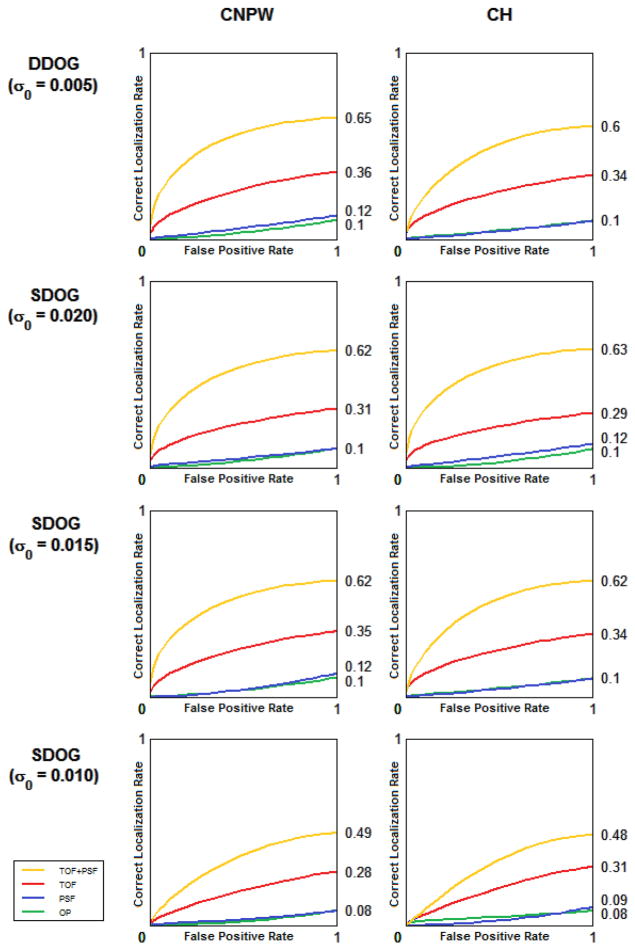
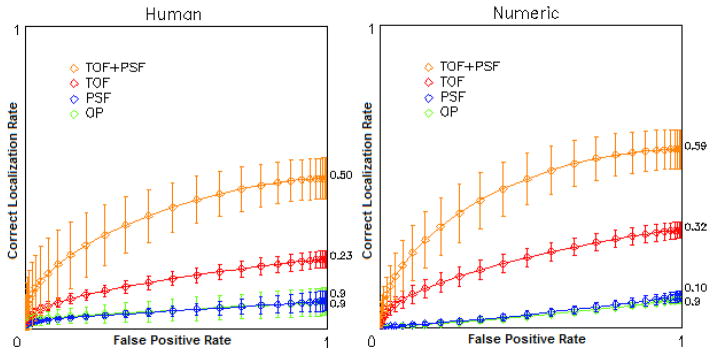
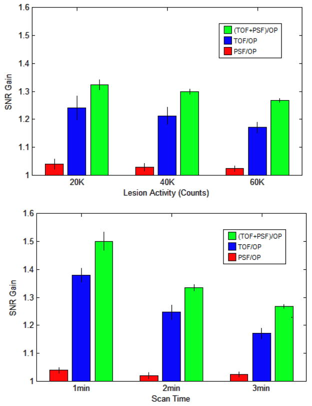
Time-of-flight (TOF) and point spread function (PSF) modeling have been shown to improve PET reconstructions, but the impact on physicians in the clinical setting has not been thoroughly investigated. A lesion detection and localization study was performed using simulated lesions in real patient images. Four reconstruction schemes were considered: ordinary Poisson OSEM (OP) alone and combined with TOF, PSF, and TOF + PSF. The images were presented to physicians experienced in reading PET images, and the performance of each was quantified using localization receiver operating characteristic. Numerical observers (non-prewhitening and Hotelling) were used to identify optimal reconstruction parameters, and observer SNR was compared to the performance of the physicians. The numerical models showed good agreement with human performance, and best performance was achieved by both when using TOF + PSF. These findings suggest a large potential benefit of TOF + PSF for oncology PET studies, especially in the detection of small, low-intensity, focal disease in larger patients.
飞行时间(TOF)和点扩散函数(PSF)建模已被证明可以改善 PET 重建,但它们对临床医生的影响尚未得到彻底研究。本研究使用真实患者图像中的模拟病变进行了病变检测和定位研究。考虑了四种重建方案:普通泊松有序子集期望最大化(OP)单独重建和结合 TOF、PSF 和 TOF+PSF 重建。将图像呈现给有阅读 PET 图像经验的医生,并使用定位接收者操作特性曲线量化每种方法的性能。使用数值观察者(非预白化和霍特林)来确定最佳重建参数,并将观察者信噪比与医生的性能进行比较。数值模型与人类表现具有很好的一致性,当使用 TOF+PSF 时,两者都能达到最佳性能。这些发现表明,TOF+PSF 对肿瘤学 PET 研究有很大的潜在益处,特别是在检测较大患者中较小、低强度、局灶性疾病方面。