The Laboratory of the Molecular Mechanisms of Hemostasis, the Center for Theoretical Problems of Physicochemical Pharmacology RAS, Moscow, Russia.
PLoS One. 2013;8(2):e55688. doi: 10.1371/journal.pone.0055688. Epub 2013 Feb 6.
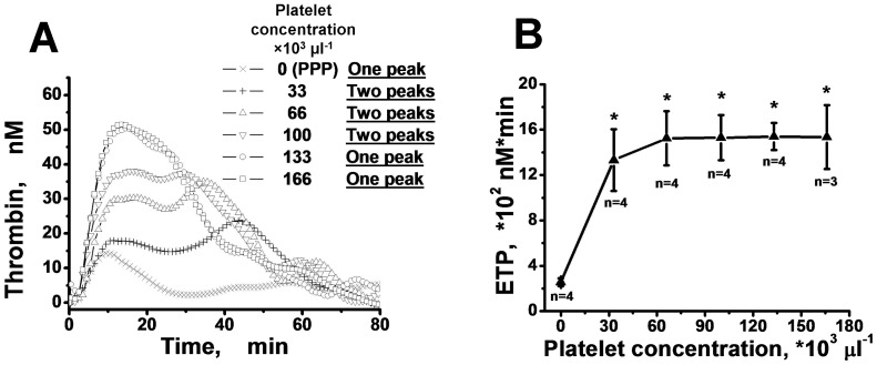
Thrombin generation assay is a convenient and widely used method for analysis of the blood coagulation system status. Thrombin generation curve (TGC) is usually bell-shaped with a single peak, but there are exceptions. In particular, TGC in platelet-rich plasma (PRP) can sometimes have two peaks.
We sought to understand the mechanism underlying the occurrence of two peaks in the PRP thrombin generation curve.
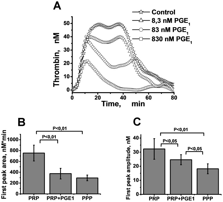
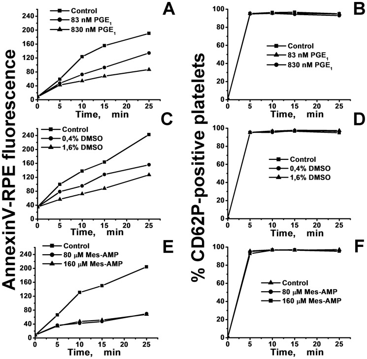
Tissue factor-induced thrombin generation in PRP and platelet-poor plasma (PPP) was monitored using continuous measurement of the hydrolysis rate of the thrombin-specific fluorogenic substrate Z-Gly-Gly-Arg-AMC. Expression of phosphatidylserine (PS) and CD62P on the surface of activated platelets was measured by flow cytometry using corresponding fluorescently labeled markers.
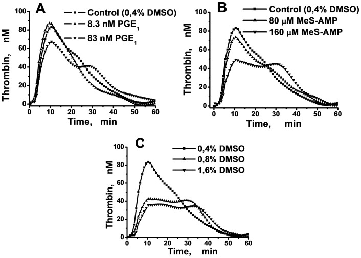
The addition of the P(2)Y(12) receptor antagonist MeS-AMP (160 µM), 83 nM prostaglandin E(1) (PGE(1)), or 1.6% DMSO to PRP caused the appearance of two peaks in the TGC. The PS exposure after thrombin activation on washed platelets in a suspension supplemented with DMSO, PGE(1) or MeS-AMP was delayed, which could indicate mechanism of the second peak formation. Supplementation of PRP with 1.6% DMSO plus 830 nM PGE(1) mediated the disappearance of the second peak and decreased the amplitude of the first peak. Increasing the platelet concentration in the PRP promoted the consolidation of the two peaks into one.
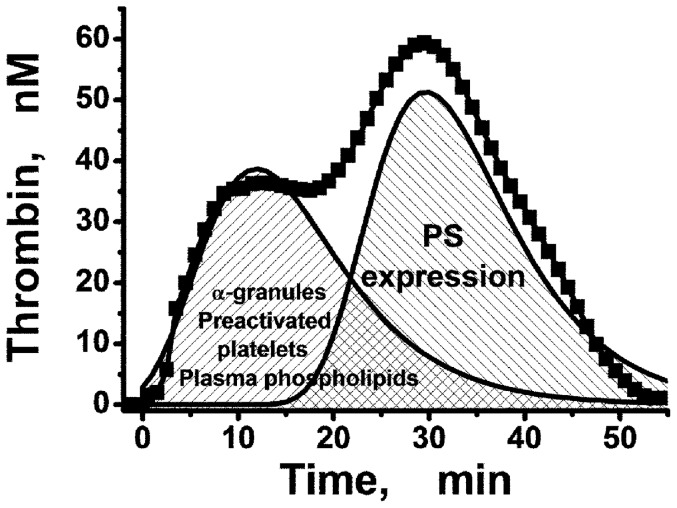
Procoagulant tenase and prothrombinase complexes in PRP assemble on phospholipid surfaces containing PS of two types--plasma lipoproteins and the surface of activated platelets. Thrombin generation in the PRP can be two-peaked. The second peak appears in the presence of platelet antagonists as a result of delayed PS expression on platelets, which leads to delayed assembly of the membrane-dependent procoagulant complexes and a second wave of thrombin generation.
凝血酶生成分析是一种方便且广泛应用的分析血液凝固系统状态的方法。凝血酶生成曲线(TGC)通常呈钟形,有单个峰值,但也有例外。特别是富含血小板的血浆(PRP)中的 TGC 有时会有两个峰值。
我们试图了解富含血小板的血浆 TGC 中出现双峰的机制。
使用连续测量凝血酶特异性荧光底物 Z-Gly-Gly-Arg-AMC 的水解速率,监测组织因子诱导的富含血小板的血浆(PRP)和血小板贫乏的血浆(PPP)中的凝血酶生成。通过使用相应的荧光标记物,通过流式细胞术测量激活血小板表面的磷脂酰丝氨酸(PS)和 CD62P 的表达。
向 PRP 中添加 P2Y12 受体拮抗剂 MeS-AMP(160µM)、83nM 前列腺素 E1(PGE1)或 1.6% DMSO 会导致 TGC 出现双峰。在添加 DMSO、PGE1 或 MeS-AMP 的悬浮液中洗涤血小板的凝血酶激活后 PS 暴露延迟,这可能表明形成第二个峰的机制。在 PRP 中补充 1.6% DMSO 加 830nM PGE1 可使第二个峰消失,并降低第一个峰的幅度。增加 PRP 中的血小板浓度可促进两个峰合并为一个峰。
PRP 中的促凝酶 tenase 和 prothrombinase 复合物在含有两种类型 PS(脂蛋白和激活血小板表面)的磷脂表面上组装。PRP 中的凝血酶生成可能呈双峰状。第二个峰出现在血小板拮抗剂存在的情况下,这是由于血小板 PS 表达延迟,导致膜依赖性促凝复合物组装延迟和第二个凝血酶生成波。