Department of Radiology and Nuclear Medicine, University Medical Center Utrecht, Utrecht, The Netherlands.
PLoS One. 2013;8(2):e55742. doi: 10.1371/journal.pone.0055742. Epub 2013 Feb 6.
After yttrium-90 ((90)Y) microsphere radioembolization (RE), evaluation of extrahepatic activity and liver dosimetry is typically performed on (90)Y Bremsstrahlung SPECT images. Since these images demonstrate a low quantitative accuracy, (90)Y PET has been suggested as an alternative. The aim of this study is to quantitatively compare SPECT and state-of-the-art PET on the ability to detect small accumulations of (90)Y and on the accuracy of liver dosimetry.
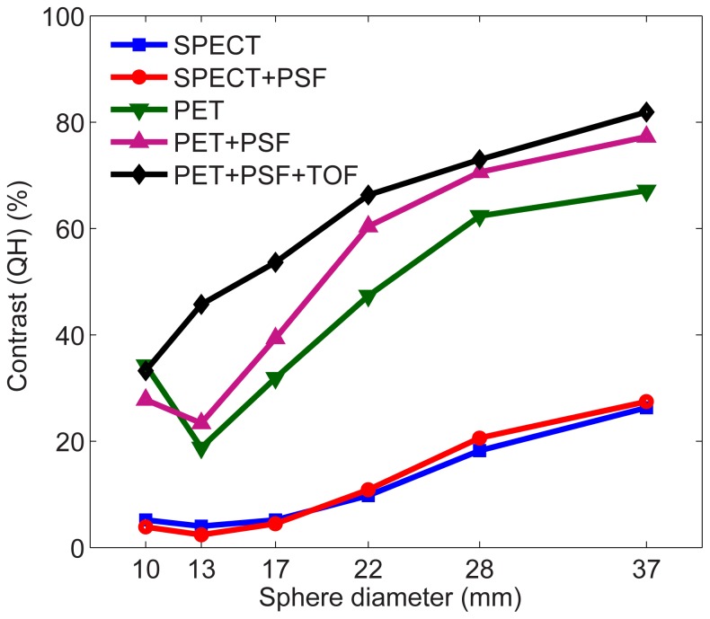
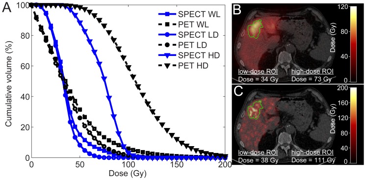
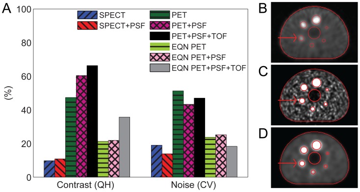
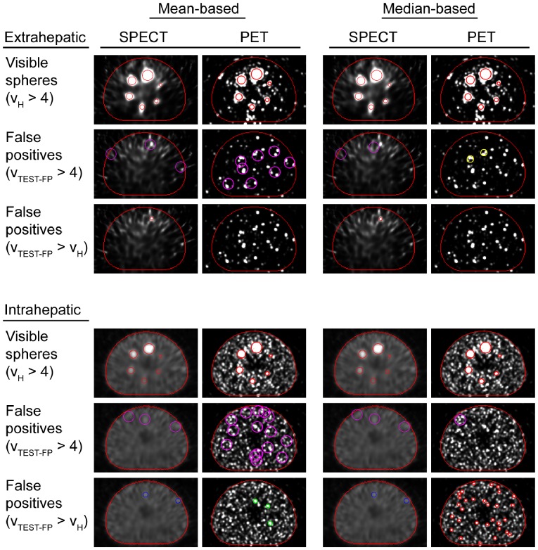
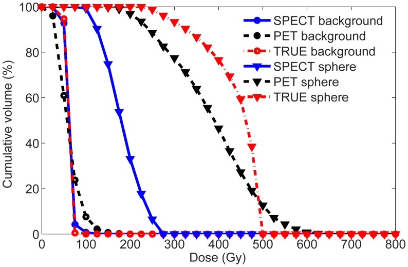
METHODOLOGY/PRINCIPAL FINDINGS: SPECT/CT and PET/CT phantom data were acquired using several acquisition and reconstruction protocols, including resolution recovery and Time-Of-Flight (TOF) PET. Image contrast and noise were compared using a torso-shaped phantom containing six hot spheres of various sizes. The ability to detect extra- and intrahepatic accumulations of activity was tested by quantitative evaluation of the visibility and unique detectability of the phantom hot spheres. Image-based dose estimates of the phantom were compared to the true dose. For clinical illustration, the SPECT and PET-based estimated liver dose distributions of five RE patients were compared. At equal noise level, PET showed higher contrast recovery coefficients than SPECT. The highest contrast recovery coefficients were obtained with TOF PET reconstruction including resolution recovery. All six spheres were consistently visible on SPECT and PET images, but PET was able to uniquely detect smaller spheres than SPECT. TOF PET-based estimates of the dose in the phantom spheres were more accurate than SPECT-based dose estimates, with underestimations ranging from 45% (10-mm sphere) to 11% (37-mm sphere) for PET, and 75% to 58% for SPECT, respectively. The differences between TOF PET and SPECT dose-estimates were supported by the patient data.
CONCLUSIONS/SIGNIFICANCE: In this study we quantitatively demonstrated that the image quality of state-of-the-art PET is superior over Bremsstrahlung SPECT for the assessment of the (90)Y microsphere distribution after radioembolization.
钇-90(90)Y 微球放射栓塞(RE)后,通常在 90Y 韧致辐射 SPECT 图像上评估肝外活性和肝脏剂量。由于这些图像显示出低定量准确性,因此已经提出 90Y PET 作为替代方法。本研究的目的是定量比较 SPECT 和最先进的 PET 在检测 90Y 小量蓄积方面的能力以及肝脏剂量计算的准确性。
方法/主要发现:使用几种采集和重建方案(包括分辨率恢复和时间飞行(TOF)PET)获得 SPECT/CT 和 PET/CT 体模数据。使用包含六个不同大小热球的躯干形状体模比较图像对比度和噪声。通过定量评估体模热球的可见性和独特可检测性来测试检测活动的肝内外蓄积的能力。将图像基剂量估计与真实剂量进行比较。为了临床说明,比较了五个 RE 患者的 SPECT 和 PET 基于的估计肝脏剂量分布。在相同噪声水平下,PET 显示出比 SPECT 更高的对比度恢复系数。使用包括分辨率恢复的 TOF PET 重建获得了最高的对比度恢复系数。所有六个球体在 SPECT 和 PET 图像上均可见,但 PET 能够比 SPECT 更独特地检测到较小的球体。TOF PET 基于的体模中剂量的估计比 SPECT 基于的剂量估计更准确,TOF PET 的低估范围为 45%(10mm 球体)至 11%(37mm 球体),SPECT 的低估范围为 75%至 58%。TOF PET 和 SPECT 剂量估计之间的差异得到了患者数据的支持。
结论/意义:在这项研究中,我们定量证明了在放射性栓塞后评估 90Y 微球分布方面,最先进的 PET 的图像质量优于韧致辐射 SPECT。