Hasegawa Akihiko, Nakahara Hiroyuki, Kinoshita Mitsuo, Asahara Hiroshi, Koziol James, Lotz Martin K
Arthritis Res Ther. 2013 Feb 14;15(1):R29. doi: 10.1186/ar4165.
Anterior cruciate ligament (ACL) degeneration is observed in most osteoarthritis (OA)-affected knee joints. However, the specific spatial and temporal relations of these changes and their association with extracellular matrix (ECM) degeneration are not well understood. The objective of this study was to characterize the patterns and relations of aging-related and OA-associated changes in ACL cells and the ECM.
Human knee joints from 80 donors (age 23 through 94) were obtained at autopsy. ACL degeneration was assessed histologically by using a quantitative scoring system. Tissue sections were analyzed for cell density, cell organization, ECM components, ECM-degrading enzymes and markers of differentiation, proliferation, and stem cells.
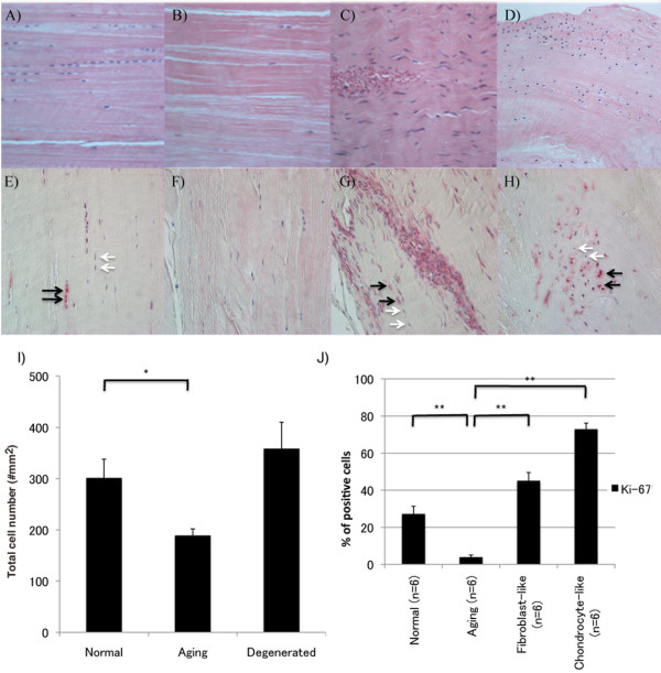
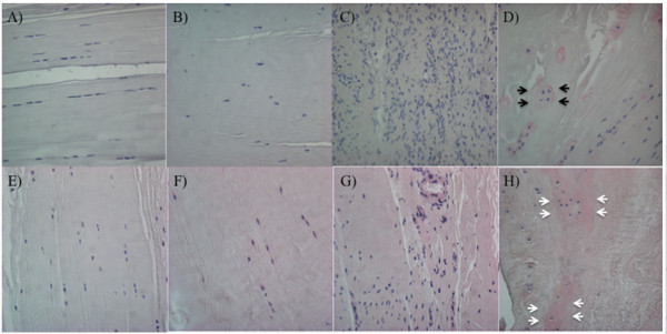
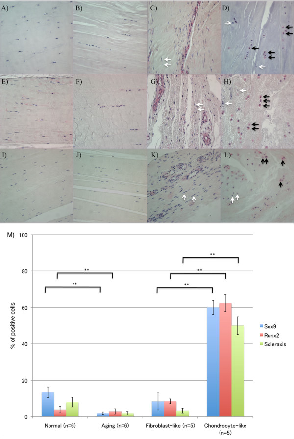
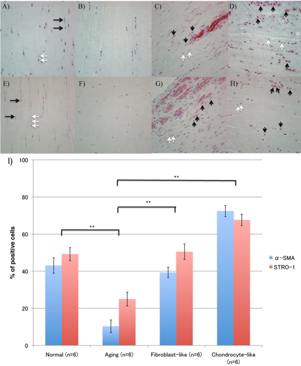
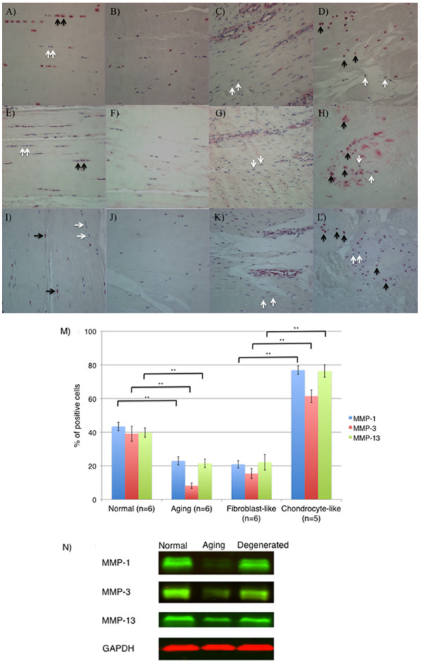
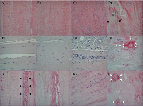
Total cell number in normal ACL decreased with aging but increased in degenerated ACL, because of the formation of perivascular cell aggregates and islands of chondrocyte-like cells. Matrix metalloproteinase (MMP)-1, -3, and -13 expression was reduced in aging ACL but increased in degenerated ACL, mainly in the chondrocyte-like cells. Collagen I was expressed throughout normal and degenerated ACL. Collagen II and X were detected only in the areas with chondroid metaplasia, which also expressed collagen III. Sox9, Runt-related transcription factor 2 (Runx2), and scleraxis expression was increased in the chondrocyte-like cells in degenerated ACL. Alpha-smooth muscle actin (α-SMA), a marker of myofibroblasts and the progenitor cell marker STRO-1, decreased with aging in normal ACL. In degenerated ACL, the new cell aggregates were positive for α-SMA and STRO-1.
ACL aging is characterized by reduced cell density and activation. In contrast, ACL degeneration is associated with cell recruitment or proliferation, including progenitor cells or myofibroblasts. Abnormally differentiated chondrocyte-like cell aggregates in degenerated ACL produce abnormal ECM and may predispose to mechanical failure.
在大多数受骨关节炎(OA)影响的膝关节中都观察到前交叉韧带(ACL)退变。然而,这些变化的具体时空关系及其与细胞外基质(ECM)退变的关联尚不清楚。本研究的目的是描述ACL细胞和ECM中与衰老相关及OA相关变化的模式和关系。
从80名捐赠者(年龄23至94岁)的膝关节在尸检时获取样本。使用定量评分系统对ACL退变进行组织学评估。对组织切片进行细胞密度、细胞组织、ECM成分、ECM降解酶以及分化、增殖和干细胞标志物的分析。
正常ACL中的细胞总数随年龄增长而减少,但在退变的ACL中增加,这是由于血管周围细胞聚集体和软骨样细胞岛的形成。基质金属蛋白酶(MMP)-1、-3和-13在衰老的ACL中表达降低,但在退变的ACL中增加,主要在软骨样细胞中。胶原蛋白I在正常和退变的ACL中均有表达。胶原蛋白II和X仅在软骨化生区域被检测到,该区域也表达胶原蛋白III。Sox9、Runx相关转录因子2(Runx2)和硬骨素在退变的ACL中的软骨样细胞中表达增加。α-平滑肌肌动蛋白(α-SMA),一种肌成纤维细胞标志物和祖细胞标志物STRO-1,在正常ACL中随年龄增长而减少。在退变的ACL中,新的细胞聚集体对α-SMA和STRO-1呈阳性。
ACL衰老的特征是细胞密度降低和激活减少。相比之下,ACL退变与细胞募集或增殖有关,包括祖细胞或肌成纤维细胞。退变的ACL中异常分化的软骨样细胞聚集体产生异常的ECM,并可能导致机械性失效。