University Hospital La Paz, Interventional Cardiology Department, Madrid, Spain.
Korean Circ J. 2013 Jan;43(1):44-7. doi: 10.4070/kcj.2013.43.1.44. Epub 2013 Jan 31.
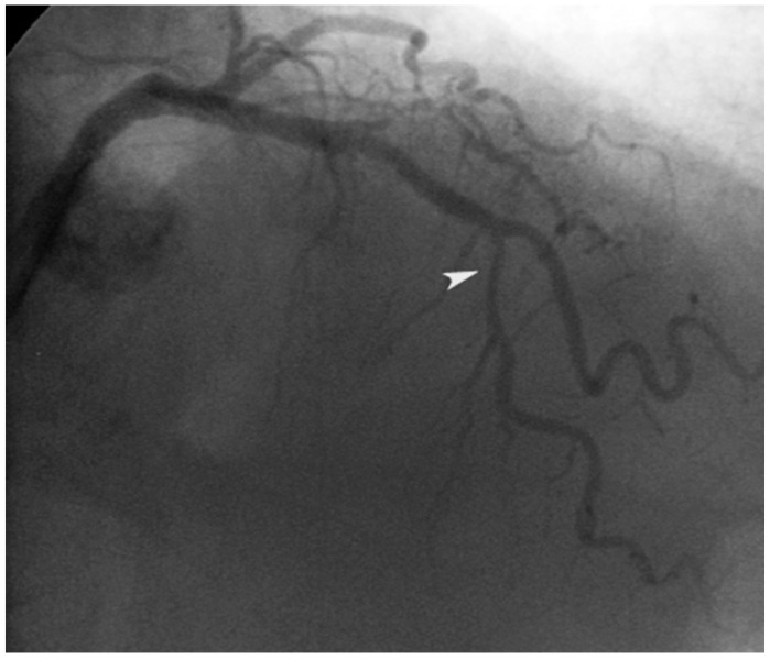
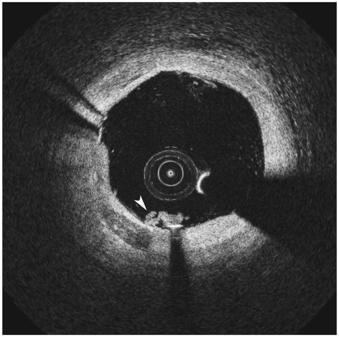
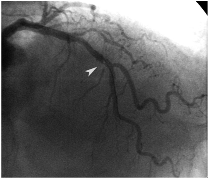
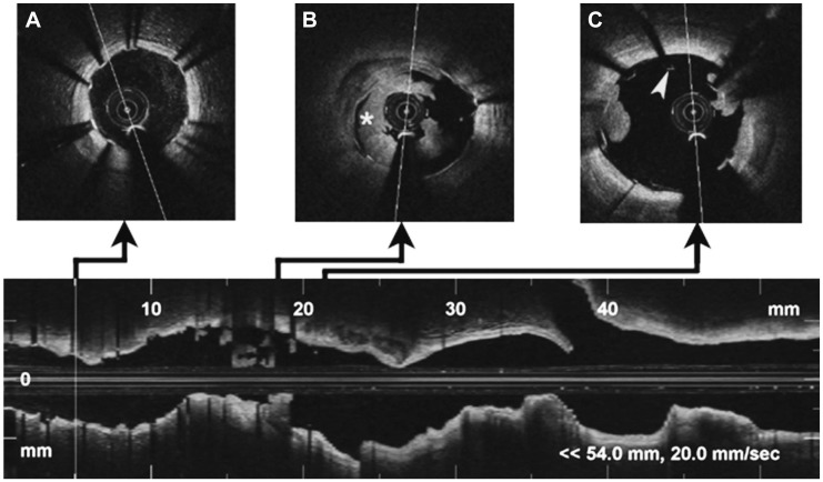
Although its use in daily practice is not common, optical coherence tomography (OCT) is a powerful research tool in invasive cardiology. This report describes a hazy angiography image after percutaneous coronary intervention that has been assessed using OCT. Based on the results of the OCT, the patient underwent an elective coronary angioplasty with standard anticoagulation. After implantation of the stent, an intracoronary hazy image was seen on angiography. The use of OCT permitted a correct diagnosis and a successful treatment. This paper provides a discussion of the advantages and disadvantages of OCT, and a comparison with intravascular ultrasound.
虽然在日常实践中并不常用,但光学相干断层扫描(OCT)是介入心脏病学中强有力的研究工具。本报告描述了经皮冠状动脉介入治疗后使用 OCT 评估的模糊血管造影图像。根据 OCT 的结果,患者接受了标准抗凝的选择性冠状动脉血管成形术。支架植入后,血管造影显示冠状动脉内出现模糊图像。OCT 的使用可以明确诊断并成功治疗。本文讨论了 OCT 的优缺点,并与血管内超声进行了比较。