Qian Zhen-Yu, Yang Ming-Feng, Zuo Ke-Qiang, Cheng Jie, Xiao Hong-Bing, Ding Wei-Xing
Department of General Surgery, Tongji Hospital, Tongji University, Shanghai 200065;
Exp Ther Med. 2013 Feb;5(2):631-635. doi: 10.3892/etm.2012.830. Epub 2012 Nov 23.
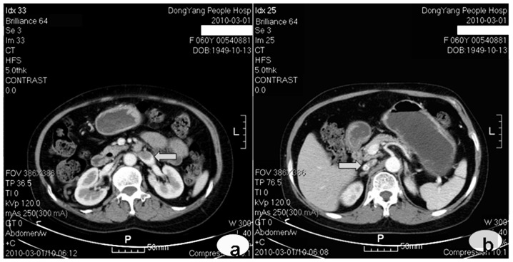
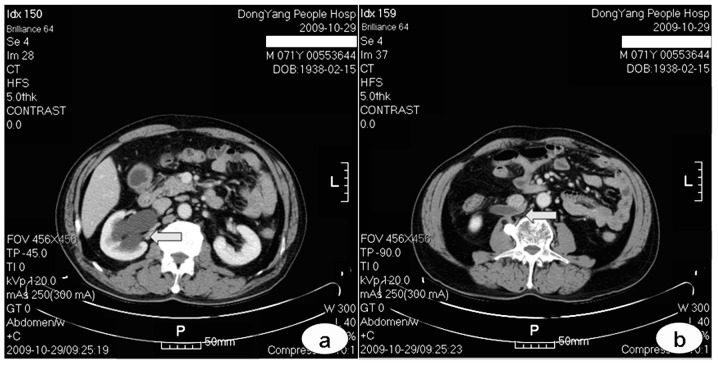
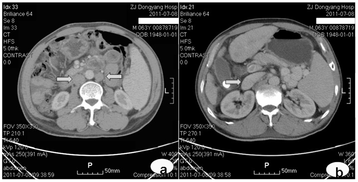
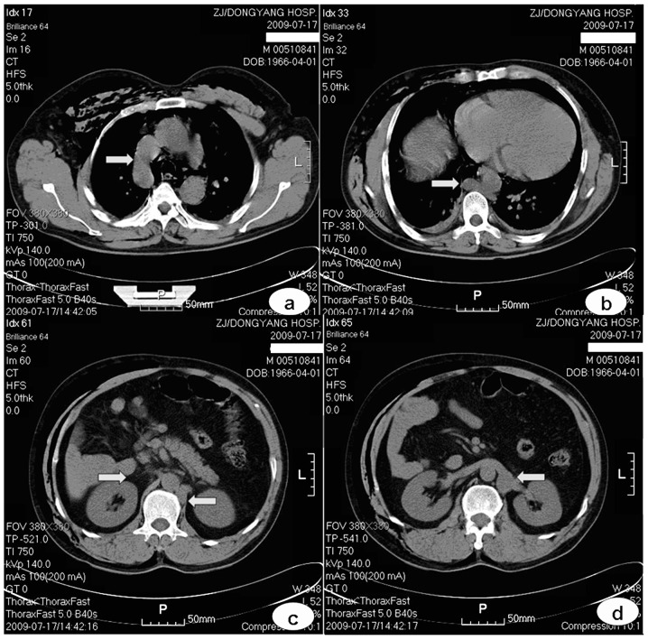
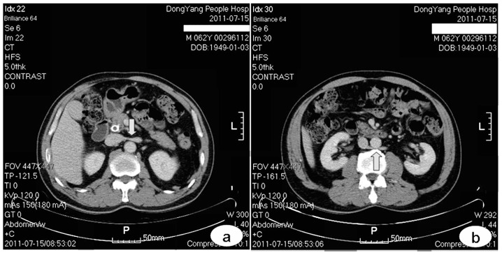
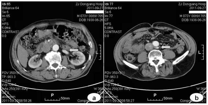
This study aimed to review and analyse the computed tomography (CT) imaging results of frequently encountered developmental anomalies of the inferior vena cava (IVC). The underlying clinical significance was evaluated with reference to the relevant literature. CT images of patients who received abdominal or thoracic scanning between July 2009 and September 2011 were reviewed. Developmental anomalies observed in the IVC were identified and categorised. Images of the cases with typical anomalies were presented and their developmental mechanism, as well as clinical significance, was discussed. The most frequently encountered IVC developmental anomalies include the left vena cava, double vena cava, azygos continuation of the IVC, left circumaortic renal vein, left retroaortic renal vein and retrocaval ureter. The embryogenesis of the IVC is a complex process that results in various congenital anomalies. The developmental anomalies of the IVC are distinguished using a CT scan and have significant implications on clinical perspective.
本研究旨在回顾和分析下腔静脉(IVC)常见发育异常的计算机断层扫描(CT)成像结果。参考相关文献评估其潜在的临床意义。回顾了2009年7月至2011年9月期间接受腹部或胸部扫描患者的CT图像。识别并分类IVC中观察到的发育异常。展示典型异常病例的图像,并讨论其发育机制以及临床意义。最常见的IVC发育异常包括左位腔静脉、双腔静脉、IVC奇静脉延续、左主动脉周围肾静脉、左主动脉后肾静脉和腔静脉后输尿管。IVC的胚胎发生是一个复杂的过程,会导致各种先天性异常。IVC的发育异常可通过CT扫描鉴别,对临床具有重要意义。