Wylie Douglas R
Centre for Neuroscience and Department of Psychology, University of Alberta Edmonton, AB, Canada.
Front Behav Neurosci. 2013 Feb 12;7:4. doi: 10.3389/fnbeh.2013.00004. eCollection 2013.
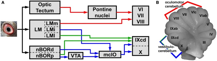
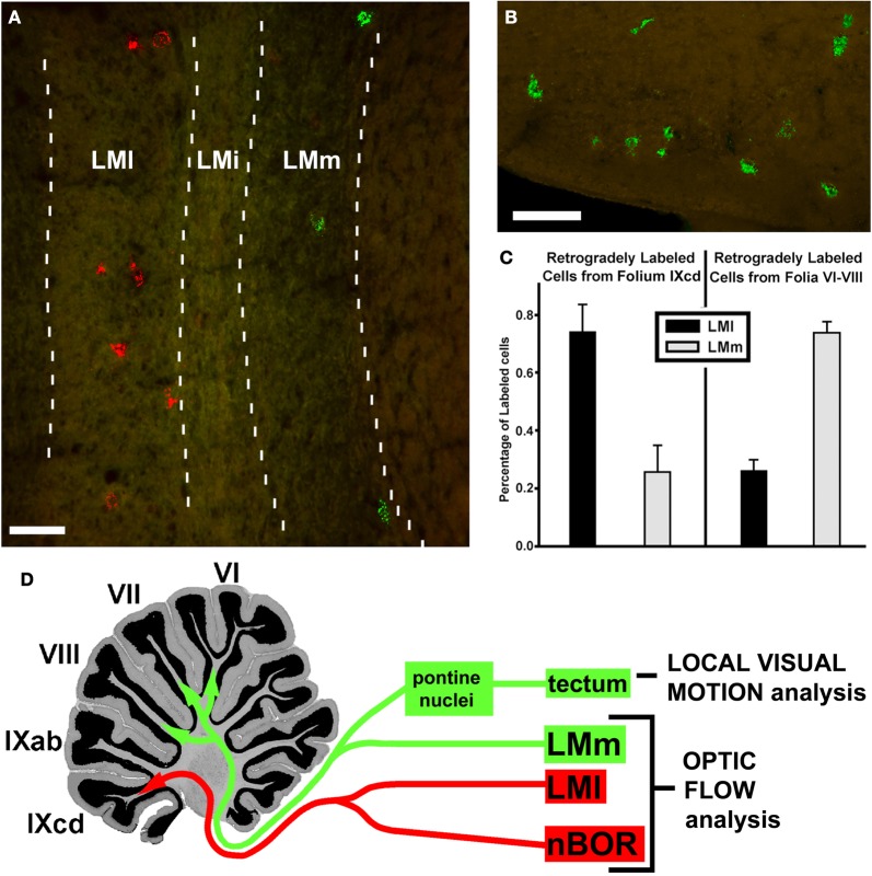
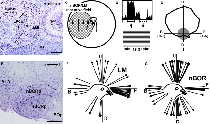
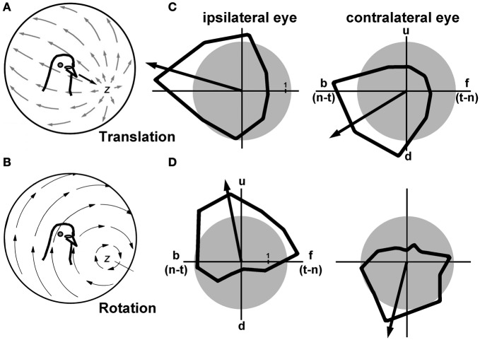
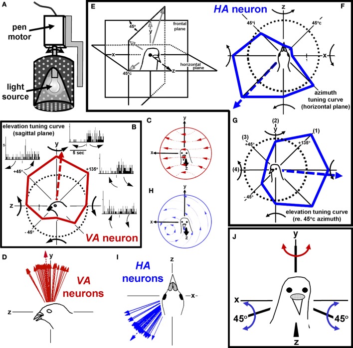
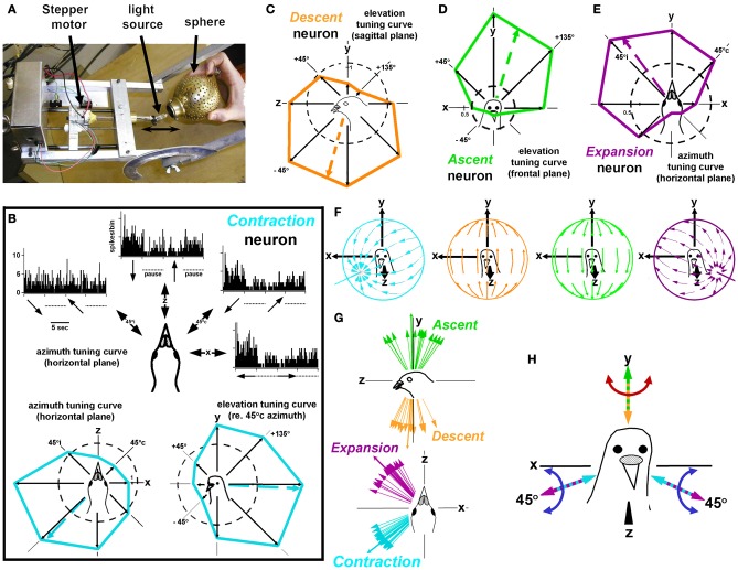
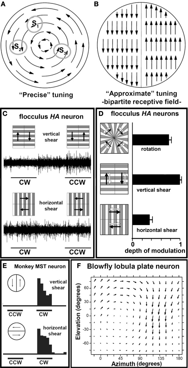
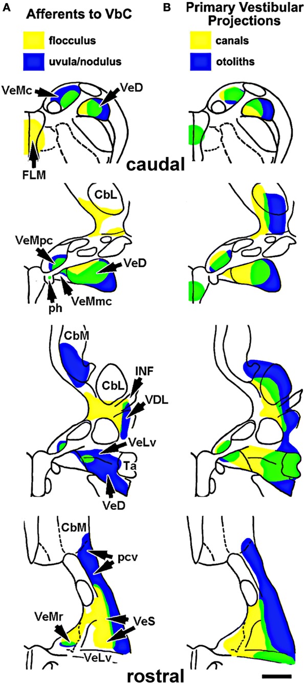
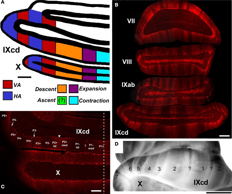
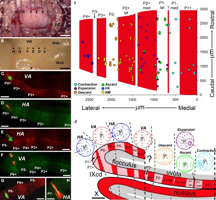
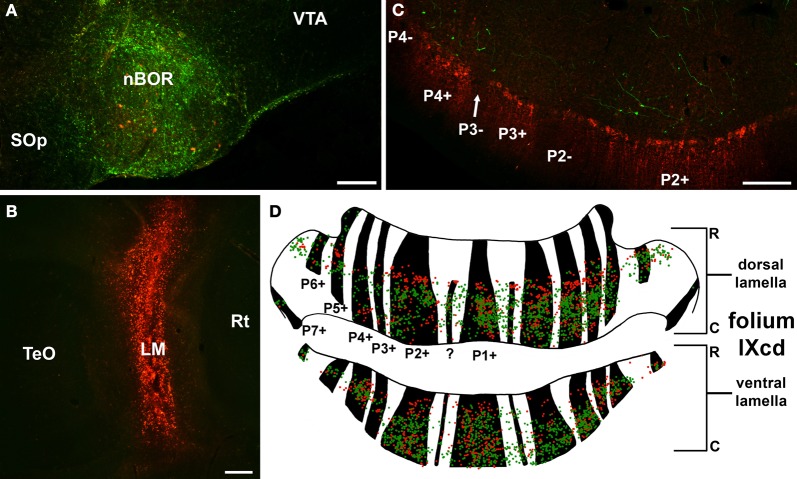
In this paper I describe the key features of optic flow processing in pigeons. Optic flow is the visual motion that occurs across the entire retina as a result of self-motion and is processed by subcortical visual pathways that project to the cerebellum. These pathways originate in two retinal-recipient nuclei, the nucleus of the basal optic root (nBOR) and the nucleus lentiformis mesencephali, which project to the vestibulocerebellum (VbC) (folia IXcd and X), directly as mossy fibers, and indirectly as climbing fibers from the inferior olive. Optic flow information is integrated with vestibular input in the VbC. There is a clear separation of function in the VbC: Purkinje cells in the flocculus process optic flow resulting from self-rotation, whereas Purkinje cells in the uvula/nodulus process optic flow resulting from self-translation. Furthermore, Purkinje cells with particular optic flow preferences are organized topographically into parasagittal "zones." These zones are correlated with expression of the isoenzyme aldolase C, also known as zebrin II (ZII). ZII expression is heterogeneous such that there are parasagittal stripes of Purkinje cells that have high expression (ZII+) alternating with stripes of Purkinje cells with low expression (ZII-). A functional zone spans a ZII± stripe pair. That is, each zone that contains Purkinje cells responsive to a particular pattern of optic flow is subdivided into a strip containing ZII+ Purkinje cells and a strip containing ZII- Purkinje cells. Additionally, there is optic flow input to folia VI-VIII of the cerebellum from lentiformis mesencephali. These folia also receive visual input from the tectofugal system via pontine nuclei. As the tectofugal system is involved in the analysis of local motion, there is integration of optic flow and local motion information in VI-VIII. This part of the cerebellum may be important for moving through a cluttered environment.
在本文中,我描述了鸽子视流处理的关键特征。视流是由于自身运动而在整个视网膜上发生的视觉运动,由投射到小脑的皮层下视觉通路进行处理。这些通路起源于两个视网膜接收核,即基底视根核(nBOR)和中脑豆状核,它们直接作为苔藓纤维投射到前庭小脑(VbC)(小叶IXcd和X),并间接作为来自下橄榄核的攀缘纤维投射到前庭小脑。视流信息在前庭小脑中与前庭输入整合。前庭小脑存在明显的功能分离:绒球中的浦肯野细胞处理由自身旋转产生的视流,而蚓垂/小结中的浦肯野细胞处理由自身平移产生的视流。此外,具有特定视流偏好的浦肯野细胞按地形组织成矢状旁“区”。这些区与同工酶醛缩酶C(也称为斑马蛋白II,ZII)的表达相关。ZII的表达是异质的,因此浦肯野细胞的矢状旁条纹中存在高表达(ZII +)与低表达(ZII -)的浦肯野细胞条纹交替出现的情况。一个功能区跨越一对ZII±条纹。也就是说,每个包含对特定视流模式有反应的浦肯野细胞的区被细分为一个包含ZII +浦肯野细胞的条带和一个包含ZII -浦肯野细胞的条带。此外,中脑豆状核有视流输入到小脑的小叶VI - VIII。这些小叶还通过脑桥核从前庭顶盖系统接收视觉输入。由于前庭顶盖系统参与局部运动分析,因此在小叶VI - VIII中存在视流和局部运动信息的整合。小脑的这一部分对于在杂乱环境中移动可能很重要。