Division of Cardiology, Department of Internal Medicine, Kaohsiung Medical University Hospital, Kaohsiung Medical University, Kaohsiung, Taiwan.
PLoS One. 2013;8(2):e55840. doi: 10.1371/journal.pone.0055840. Epub 2013 Feb 7.
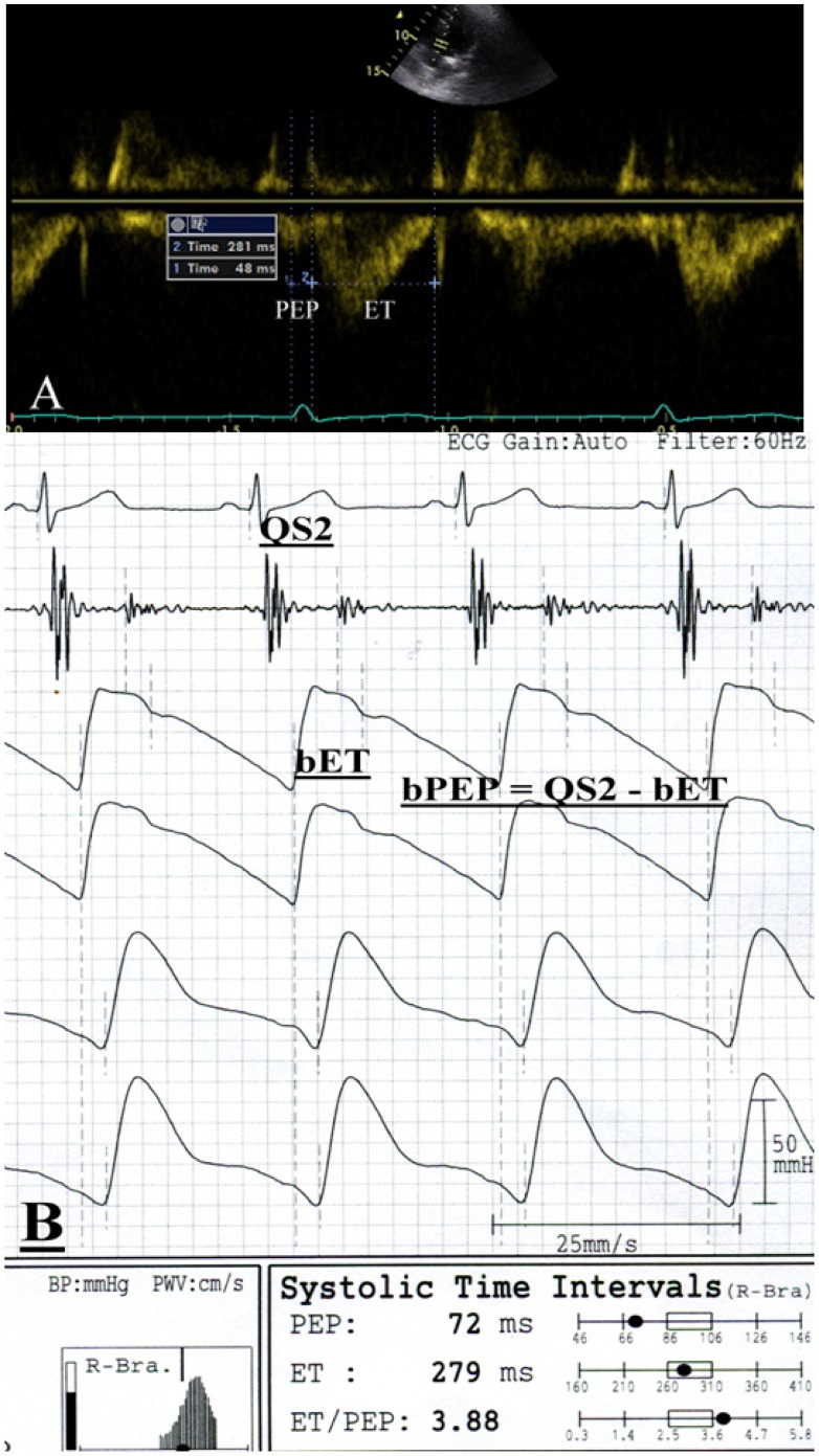
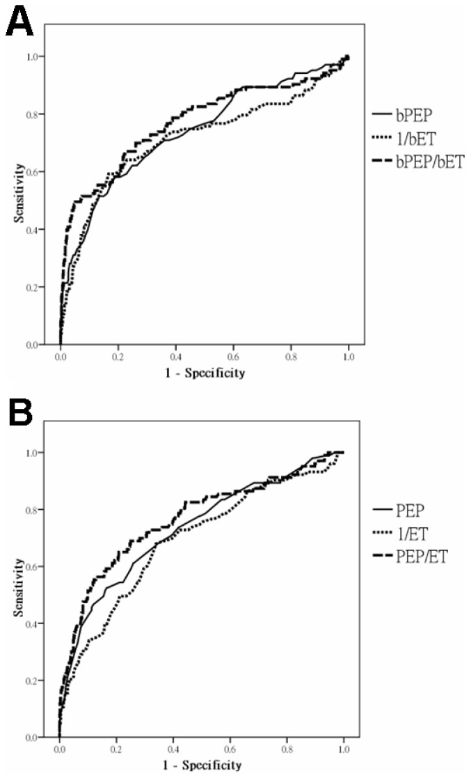
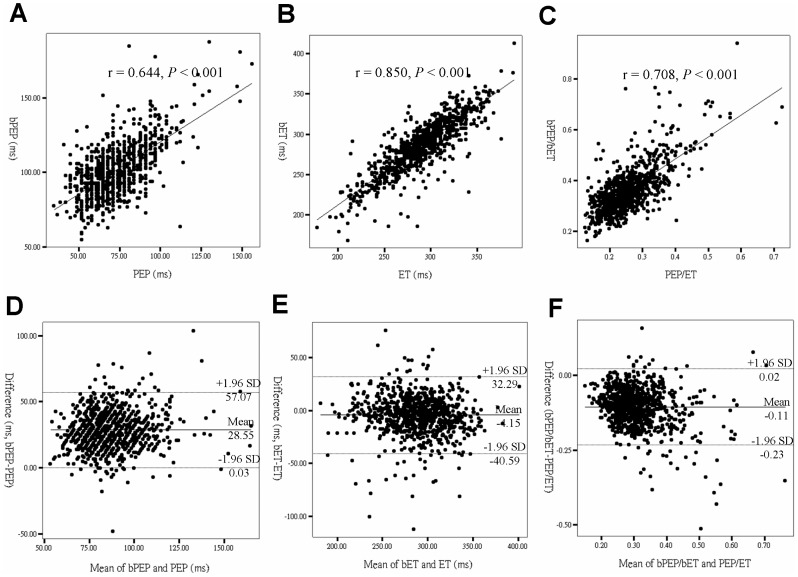
Systolic time interval (STI) is an established noninvasive technique for the assessment of cardiac function. Brachial STIs can be automatically determined by an ankle-brachial index (ABI)-form device. The aims of this study are to evaluate whether the STIs measured from ABI-form device can represent those measured from echocardiography and to compare the diagnostic values of brachial and echocardiographic STIs in the prediction of left ventricular ejection fraction (LVEF) <50%. A total of 849 patients were included in the study. Brachial pre-ejection period (bPEP) and brachial ejection time (bET) were measured using an ABI-form device and pre-ejection period (PEP) and ejection time (ET) were measured from echocardiography. Agreement was assessed by correlation coefficient and Bland-Altman plot. Brachial STIs had a significant correlation with echocardiographic STIs (r = 0.644, P<0.001 for bPEP and PEP; r = 0.850, P<0.001 for bET and ET; r = 0.708, P<0.001 for bPEP/bET and PEP/ET). The disagreement between brachial and echocardiographic STIs (brachial STIs minus echocardiographic STIs) was 28.55 ms for bPEP and PEP, -4.15 ms for bET and ET and -0.11 for bPEP/bET and PEP/ET. The areas under the curve for bPEP/bET and PEP/ET in the prediction of LVEF <50% were 0.771 and 0.765, respectively. Brachial STIs were good alternatives to STIs obtained from echocardiography and also helpful in prediction of LVEF <50%. Brachial STIs automatically obtained from an ABI-form device may be helpful for evaluation of left ventricular systolic dysfunction.
心动周期时间间隔(STI)是评估心功能的一种既定的非侵入性技术。通过踝臂指数(ABI)形式的设备可以自动确定肱动脉 STI。本研究的目的是评估从 ABI 形式的设备测量的 STI 是否可以代表从超声心动图测量的 STI,并比较肱动脉和超声心动图 STI 在预测左心室射血分数(LVEF)<50%中的诊断价值。共有 849 名患者纳入研究。使用 ABI 形式的设备测量肱动脉射前期(bPEP)和肱动脉射血时间(bET),并从超声心动图测量射前期(PEP)和射血时间(ET)。通过相关系数和 Bland-Altman 图评估一致性。肱动脉 STI 与超声心动图 STI 有显著相关性(bPEP 和 PEP 之间的 r 值为 0.644,P<0.001;bET 和 ET 之间的 r 值为 0.850,P<0.001;bPEP/bET 和 PEP/ET 之间的 r 值为 0.708,P<0.001)。肱动脉 STI 与超声心动图 STI 之间的差异(肱动脉 STI 减去超声心动图 STI)为 bPEP 和 PEP 为 28.55 ms,bET 和 ET 为-4.15 ms,bPEP/bET 和 PEP/ET 为-0.11。bPEP/bET 和 PEP/ET 在预测 LVEF<50%中的曲线下面积分别为 0.771 和 0.765。肱动脉 STI 是超声心动图获得的 STI 的良好替代物,对预测 LVEF<50%也有帮助。从 ABI 形式的设备自动获得的肱动脉 STI 可能有助于评估左心室收缩功能障碍。