1st Institute of Pathology and Experimental Cancer Research, Semmelweis University, Budapest, Hungary.
J Transl Med. 2013 Feb 18;11:43. doi: 10.1186/1479-5876-11-43.
Besides being a preferential site of early metastasis, the sentinel lymph node (SLN) is also a privileged site of T-cell priming, and may thus be an appropriate target for investigating cell types involved in antitumor immune reactions.
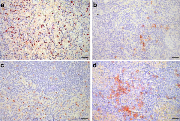
In this retrospective study we determined the prevalence of OX40+ activated T lymphocytes, FOXP3+ (forkhead box P3) regulatory T cells, DC-LAMP+ (dendritic cell-lysosomal associated membrane protein) mature dendritic cells (DCs) and CD123+ plasmacytoid DCs by immunohistochemistry in 100 SLNs from 60 melanoma patients. Density values of each cell type in SLNs were compared to those in non-sentinel nodes obtained from block dissections (n = 37), and analyzed with regard to associations with clinicopathological parameters and disease outcome.
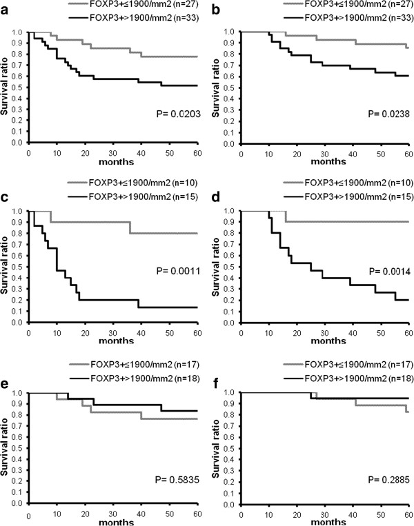
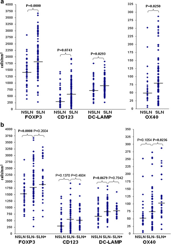
Sentinel nodes showed elevated amount of all cell types studied in comparison to non-sentinel nodes. Metastatic SLNs had higher density of OX40+ lymphocytes compared to tumor-negative nodes, while no significant difference was observed in the case of the other cell types studied. In patients with positive sentinel node status, high amount of FOXP3+ cells in SLNs was associated with shorter progression-free (P = 0.0011) and overall survival (P = 0.0014), while no significant correlation was found in the case of sentinel-negative patients. The density of OX40+, CD123+ or DC-LAMP+ cells did not show significant association with the outcome of the disease.
Taken together, our results are compatible with the hypothesis of functional competence of sentinel lymph nodes based on the prevalence of the studied immune cells. The density of FOXP3+ lymphocytes showed association with progression and survival in patients with positive SLN status, while the other immune markers studied did not prove of prognostic importance. These results, together with our previous findings on the prognostic value of activated T cells and mature DCs infiltrating primary melanomas, suggest that immune activation-associated markers in the primary tumor may have a higher impact than those in SLNs on the prognosis of the patients. On the other hand, FOXP3+ cell density in SLNs, but not in the primary tumor, was found predictive of disease outcome in melanoma patients.
除了是早期转移的首选部位外,前哨淋巴结 (SLN) 也是 T 细胞启动的特权部位,因此可能是研究参与抗肿瘤免疫反应的细胞类型的合适目标。
在这项回顾性研究中,我们通过免疫组织化学方法检测了 60 例黑色素瘤患者的 100 个 SLN 中 OX40+激活的 T 淋巴细胞、FOXP3+(叉头框 P3)调节性 T 细胞、DC-LAMP+(树突状细胞-溶酶体相关膜蛋白)成熟树突状细胞 (DC) 和 CD123+浆细胞样 DC 的患病率。将 SLN 中每种细胞类型的密度值与从块解剖获得的非前哨淋巴结(n=37)进行比较,并分析其与临床病理参数和疾病结局的关系。
与非前哨淋巴结相比,SLN 中研究的所有细胞类型的数量均升高。与肿瘤阴性淋巴结相比,转移性 SLN 中 OX40+淋巴细胞的密度更高,而研究的其他细胞类型则没有明显差异。在前哨淋巴结阳性的患者中,SLN 中大量 FOXP3+细胞与无进展(P=0.0011)和总生存期(P=0.0014)较短相关,而在前哨淋巴结阴性的患者中未发现显著相关性。OX40+、CD123+或 DC-LAMP+细胞的密度与疾病结局无显著相关性。
总的来说,我们的结果与基于研究免疫细胞流行率的前哨淋巴结功能能力的假设一致。FOXP3+淋巴细胞的密度与 SLN 阳性患者的进展和生存相关,而研究的其他免疫标志物则没有证明具有预后意义。这些结果,以及我们之前关于原发性黑色素瘤中激活的 T 细胞和成熟 DC 浸润的预后价值的发现,表明原发性肿瘤中与免疫激活相关的标志物对患者的预后可能比 SLN 中的标志物具有更大的影响。另一方面,仅在 SLN 中而不在原发性肿瘤中发现 FOXP3+细胞密度可预测黑色素瘤患者的疾病结局。