• 文献检索
  • 文档翻译
  • 深度研究
  • 学术资讯
  • Suppr Zotero 插件Zotero 插件
  • 邀请有礼
  • 套餐&价格
  • 历史记录
应用&插件
Suppr Zotero 插件Zotero 插件浏览器插件Mac 客户端Windows 客户端微信小程序
定价
高级版会员购买积分包购买API积分包
服务
文献检索文档翻译深度研究API 文档MCP 服务
关于我们
关于 Suppr公司介绍联系我们用户协议隐私条款
关注我们

Suppr 超能文献

核心技术专利:CN118964589B侵权必究
粤ICP备2023148730 号-1Suppr @ 2026

文献检索

告别复杂PubMed语法,用中文像聊天一样搜索,搜遍4000万医学文献。AI智能推荐,让科研检索更轻松。

立即免费搜索

文件翻译

保留排版,准确专业,支持PDF/Word/PPT等文件格式,支持 12+语言互译。

免费翻译文档

深度研究

AI帮你快速写综述,25分钟生成高质量综述,智能提取关键信息,辅助科研写作。

立即免费体验

朊病毒蛋白斑块中的铜含量低,锰含量高。

Low copper and high manganese levels in prion protein plaques.

机构信息

USGS National Wildlife Health Center, Madison, WI 53711, USA.

出版信息

Viruses. 2013 Feb 11;5(2):654-62. doi: 10.3390/v5020654.

DOI:10.3390/v5020654
PMID:23435237
原文链接:https://pmc.ncbi.nlm.nih.gov/articles/PMC3640519/
Abstract

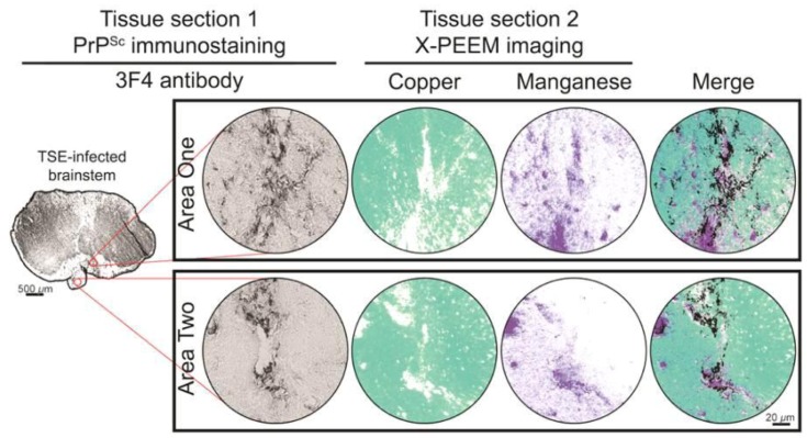
Accumulation of aggregates rich in an abnormally folded form of the prion protein characterize the neurodegeneration caused by transmissible spongiform encephalopathies (TSEs). The molecular triggers of plaque formation and neurodegeneration remain unknown, but analyses of TSE-infected brain homogenates and preparations enriched for abnormal prion protein suggest that reduced levels of copper and increased levels of manganese are associated with disease. The objectives of this study were to: (1) assess copper and manganese levels in healthy and TSE-infected Syrian hamster brain homogenates; (2) determine if the distribution of these metals can be mapped in TSE-infected brain tissue using X-ray photoelectron emission microscopy (X-PEEM) with synchrotron radiation; and (3) use X-PEEM to assess the relative amounts of copper and manganese in prion plaques in situ. In agreement with studies of other TSEs and species, we found reduced brain levels of copper and increased levels of manganese associated with disease in our hamster model. We also found that the in situ levels of these metals in brainstem were sufficient to image by X-PEEM. Using immunolabeled prion plaques in directly adjacent tissue sections to identify regions to image by X-PEEM, we found a statistically significant relationship of copper-manganese dysregulation in prion plaques: copper was depleted whereas manganese was enriched. These data provide evidence for prion plaques altering local transition metal distribution in the TSE-infected central nervous system.

摘要

富含异常折叠形式朊病毒蛋白的聚集物的积累是可传播海绵状脑病(TSEs)引起的神经退行性变的特征。斑块形成和神经退行性变的分子触发因素仍不清楚,但对 TSE 感染的脑匀浆和富含异常朊病毒蛋白的制剂的分析表明,铜水平降低和锰水平升高与疾病有关。本研究的目的是:(1)评估健康和 TSE 感染的叙利亚仓鼠脑匀浆中的铜和锰水平;(2)确定使用同步辐射的 X 射线光电子发射显微镜(X-PEEM)是否可以在 TSE 感染的脑组织中绘制这些金属的分布;(3)使用 X-PEEM 评估原位朊病毒斑块中铜和锰的相对含量。与其他 TSE 和物种的研究一致,我们发现我们的仓鼠模型中与疾病相关的铜脑水平降低和锰水平升高。我们还发现,脑桥中这些金属的原位水平足以通过 X-PEEM 成像。通过使用免疫标记的朊病毒斑块在直接相邻的组织切片中识别要通过 X-PEEM 成像的区域,我们发现朊病毒斑块中铜-锰失调存在统计学上的显著关系:铜耗竭而锰富集。这些数据为朊病毒斑块改变 TSE 感染中枢神经系统中局部过渡金属分布提供了证据。

https://cdn.ncbi.nlm.nih.gov/pmc/blobs/b4c0/3640519/13eb032d3328/viruses-05-00654-g001.jpg

相似文献

1
Low copper and high manganese levels in prion protein plaques.朊病毒蛋白斑块中的铜含量低,锰含量高。
Viruses. 2013 Feb 11;5(2):654-62. doi: 10.3390/v5020654.
2
Does an infrasonic acoustic shock wave resonance of the manganese 3+ loaded/copper depleted prion protein initiate the pathogenesis of TSE?负载锰3+ / 铜缺乏的朊病毒蛋白的次声冲击波共振会引发传染性海绵状脑病的发病机制吗?
Med Hypotheses. 2003 Jun;60(6):797-820. doi: 10.1016/s0306-9877(03)00007-0.
3
Does an ultra violet photooxidation of the manganese-loaded/copper-depleted prion protein in the retina initiate the pathogenesis of TSE?视网膜中锰负载/铜缺失的朊病毒蛋白的紫外线光氧化是否引发了传染性海绵状脑病的发病机制?
Med Hypotheses. 2001 Jul;57(1):29-45. doi: 10.1054/mehy.2001.1305.
4
Trace elements and prion diseases: a review of the interactions of copper, manganese and zinc with the prion protein.微量元素与朊病毒疾病:铜、锰和锌与朊病毒蛋白相互作用的综述
Anim Health Res Rev. 2006 Jun-Dec;7(1-2):97-105. doi: 10.1017/S1466252307001181.
5
Prion infection impairs copper binding of cultured cells.朊病毒感染会损害培养细胞的铜结合能力。
J Biol Chem. 2003 Apr 25;278(17):14595-8. doi: 10.1074/jbc.C300092200. Epub 2003 Mar 10.
6
In situ formation of protease-resistant prion protein in transmissible spongiform encephalopathy-infected brain slices.在传染性海绵状脑病感染的脑切片中原位形成抗蛋白酶朊病毒蛋白。
J Biol Chem. 1997 Jun 13;272(24):15227-31. doi: 10.1074/jbc.272.24.15227.
7
The effects of prion protein expression on metal metabolism.朊病毒蛋白表达对金属代谢的影响。
Mol Cell Neurosci. 2009 Jun;41(2):135-47. doi: 10.1016/j.mcn.2009.02.002. Epub 2009 Feb 21.
8
Prions and manganese: A maddening beast.朊病毒与锰:令人疯狂的结合。
Metallomics. 2011 Mar;3(3):229-38. doi: 10.1039/c0mt00047g. Epub 2010 Oct 29.
9
Decreased brain copper due to copper deficiency has no effect on bovine prion proteins.铜缺乏导致的脑铜减少对牛朊蛋白没有影响。
Biochem Biophys Res Commun. 2007 Jan 26;352(4):884-8. doi: 10.1016/j.bbrc.2006.11.108. Epub 2006 Dec 1.
10
Metal imbalance and compromised antioxidant function are early changes in prion disease.金属失衡和抗氧化功能受损是朊病毒疾病的早期变化。
Biochem J. 2002 Feb 15;362(Pt 1):253-8. doi: 10.1042/0264-6021:3620253.

引用本文的文献

1
Copper drives prion protein phase separation and modulates aggregation.铜离子驱动朊病毒蛋白相分离并调节聚集。
Sci Adv. 2023 Nov 3;9(44):eadi7347. doi: 10.1126/sciadv.adi7347.
2
Movement of Chronic Wasting Disease Prions in Prairie, Boreal and Alpine Soils.慢性消耗病朊病毒在草原、北方森林和高山土壤中的移动
Pathogens. 2023 Feb 7;12(2):269. doi: 10.3390/pathogens12020269.
3
Neurometals in the Pathogenesis of Prion Diseases.神经金属在朊病毒病发病机制中的作用。

本文引用的文献

1
Prion protein facilitates uptake of zinc into neuronal cells.朊病毒蛋白促进锌进入神经元细胞。
Nat Commun. 2012;3:1134. doi: 10.1038/ncomms2135.
2
Prions.朊病毒。
Cold Spring Harb Perspect Biol. 2011 Jan 1;3(1):a006833. doi: 10.1101/cshperspect.a006833.
3
Prions and manganese: A maddening beast.朊病毒与锰:令人疯狂的结合。
Int J Mol Sci. 2021 Jan 28;22(3):1267. doi: 10.3390/ijms22031267.
4
The Effects of Divalent Cation-Chelated Prion Fibrils on the Immune Response of EOC 13.31 Microglia Cells.二价阳离子螯合朊病毒纤维对 EOC 13.31 小神经胶质细胞免疫反应的影响。
Cells. 2020 Oct 13;9(10):2285. doi: 10.3390/cells9102285.
5
Potential role of soil properties in the spread of CWD in western Canada.土壤特性在加拿大西部慢性消耗病传播中的潜在作用。
Prion. 2014 Jan-Feb;8(1):92-9. doi: 10.4161/pri.28467.
Metallomics. 2011 Mar;3(3):229-38. doi: 10.1039/c0mt00047g. Epub 2010 Oct 29.
4
Prion protein expression level alters regional copper, iron and zinc content in the mouse brain.朊蛋白表达水平改变了小鼠脑内的区域铜、铁和锌含量。
Metallomics. 2011 Feb;3(2):206-14. doi: 10.1039/c0mt00037j. Epub 2011 Jan 24.
5
Manganese chelation therapy extends survival in a mouse model of M1000 prion disease.锰螯合疗法延长 M1000 朊病毒病小鼠模型的存活时间。
J Neurochem. 2010 Jul;114(2):440-51. doi: 10.1111/j.1471-4159.2010.06771.x. Epub 2010 Apr 23.
6
Consequences of dietary manganese and copper imbalance on neuronal apoptosis in a murine model of scrapie.饮食锰和铜失衡对朊病毒病小鼠模型中神经元凋亡的影响。
Neuropathol Appl Neurobiol. 2010 Jun;36(4):300-11. doi: 10.1111/j.1365-2990.2010.01065.x. Epub 2010 Jan 7.
7
Role of copper and manganese in prion disease progression.铜和锰在朊病毒疾病进展中的作用。
Brain Res. 2009 Oct 6;1292:155-64. doi: 10.1016/j.brainres.2009.07.051. Epub 2009 Jul 25.
8
Ultraviolet-ozone treatment reduces levels of disease-associated prion protein and prion infectivity.紫外线-臭氧处理可降低与疾病相关的朊病毒蛋白水平和朊病毒传染性。
BMC Res Notes. 2009 Jul 6;2:121. doi: 10.1186/1756-0500-2-121.
9
The effect of metal imbalances on scrapie neurodegeneration.金属失衡对羊瘙痒病神经退行性变的影响。
Zoonoses Public Health. 2010 Aug;57(5):358-66. doi: 10.1111/j.1863-2378.2009.01230.x. Epub 2009 Apr 8.
10
Abnormal brain iron homeostasis in human and animal prion disorders.人类和动物朊病毒疾病中的脑铁稳态异常。
PLoS Pathog. 2009 Mar;5(3):e1000336. doi: 10.1371/journal.ppat.1000336. Epub 2009 Mar 13.