Fan Tingjun, Ma Xiya, Zhao Jun, Wen Qian, Hu Xiuzhong, Yu Haoze, Shi Weiyun
Key Laboratory for Corneal Tissue Engineering, Ocean University of China, Qingdao, Shandong Province, China.
Mol Vis. 2013;19:400-7. Epub 2013 Feb 18.
To evaluate the performance of reconstructed tissue-engineered human corneal endothelium (TE-HCE) by corneal transplantation in cat models.
TE-HCE reconstruction was performed by culturing 1,1'-dioctadecyl-3,3,3',3'-tetramethylindocarbocyanine perchlorate (DiI)-labeled monoclonal HCE cells on denuded amniotic membranes (dAMs) in 20% fetal bovine serum-containing Dulbecco's Modified Eagle's Medium/Ham's Nutrient Mixture F12 (1:1) medium and 5% CO(2) at 37 ° C on a 24-well culture plate. The reconstructed TE-HCE was transplanted into cat corneas via lamellar keratoplasty with all of the endothelium and part of Descemet's membrane stripped. Postsurgical corneas were monitored daily with their histological properties examined during a period of 104 days after transplantation.
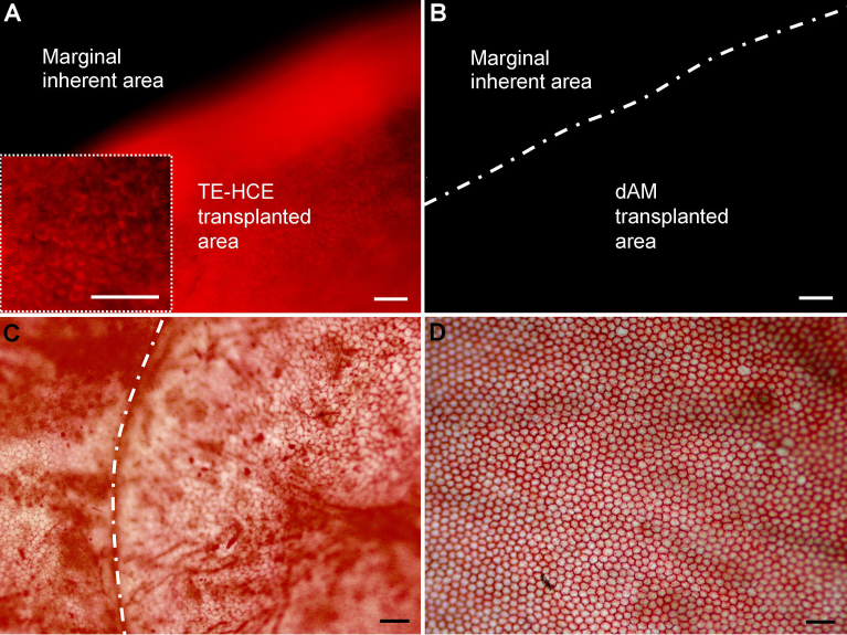
The reconstructed TE-HCE at a density of 3,413.33 ± 111.23 cells/mm(2) in average established intense cell-cell and cell-dAM junctions. After lamellar keratoplasty surgery, no obvious edema was found in TE-HCE-transplanted cat corneas, which were transparent throughout the monitoring period. In contrast, intense corneal edema developed in dAM-transplanted cat corneas, which were turbid. The corneal thickness gradually decreased to 751.33 ± 11.37 μm on day 104 after TE-HCE transplantation, while that of dAM eye was over 1,000 μm in thickness during the monitoring period. A monolayer of endothelium consisting of TE-HCE-originated cells at a density of 2,573.33 ± 0.59 cells/mm(2) attached tightly to the surface of remnant Descemet's membrane over 104 days; this was similar to the normal eye control in cell density.
The reconstructed TE-HCE was able to function as a corneal endothelium equivalent and restore corneal function in cat models.
通过在猫模型中进行角膜移植来评估重建的组织工程化人角膜内皮(TE-HCE)的性能。
通过在含20%胎牛血清的杜氏改良 Eagle 培养基/哈姆氏营养混合物 F12(1:1)培养基中,于37°C、5%二氧化碳条件下,在24孔培养板上的无细胞羊膜(dAM)上培养1,1'-二辛基-3,3,3',3'-四甲基吲哚羰花青高氯酸盐(DiI)标记的单克隆角膜内皮细胞来进行TE-HCE重建。将重建的TE-HCE通过板层角膜移植术移植到猫角膜中,去除所有内皮和部分Descemet膜。术后每天监测角膜,并在移植后104天内检查其组织学特性。
重建的TE-HCE平均密度为3413.33±111.23个细胞/mm²,形成了紧密的细胞-细胞和细胞-dAM连接。板层角膜移植术后,TE-HCE移植的猫角膜未发现明显水肿,在整个监测期内均保持透明。相比之下,dAM移植的猫角膜出现严重角膜水肿,角膜浑浊。TE-HCE移植后第104天,角膜厚度逐渐降至751.33±11.37μm,而dAM移植眼在监测期内角膜厚度超过1000μm。在104天内,由TE-HCE来源的细胞组成的单层内皮以2573.33±0.59个细胞/mm²的密度紧密附着在残余Descemet膜表面;这在细胞密度上与正常眼对照相似。
重建的TE-HCE能够在猫模型中发挥角膜内皮等效物的功能并恢复角膜功能。