Ammar David A, Kahook Malik Y
University of Colorado Hospital Eye Center, Department of Ophthalmology, University of Colorado Denver, Aurora, CO 80045, USA.
Mol Vis. 2013;19:424-9. Epub 2013 Feb 20.
Photodynamic therapy (PDT) laser light in conjunction with the benzoporphyrin derivative verteporfin is a current clinical treatment for choroidal vascular diseases such as age-related macular degeneration. The aim of this study was to examine the effects of PDT laser-activated and inactive verteporfin on various cultured ocular cells.
Primary human scleral fibroblasts (hFibro), primary human trabecular meshwork (TM) cells (hTMC), primary porcine TM cells (pTMC), and a human retinal pigment epithelial cell line (ARPE-19 cells) were treated with verteporfin with and without activation by PDT laser. Cell viability was determined according to mitochondrial enzyme activity (3-(4,5- dimethyl-2-thiazoyl)-2,5-diphenyl-2H-tetrazolium bromide assay).
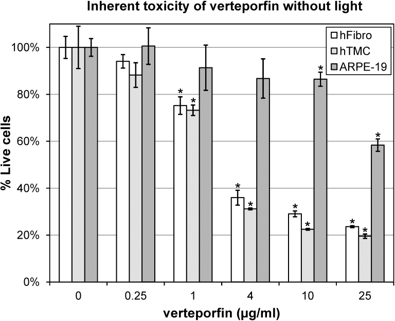
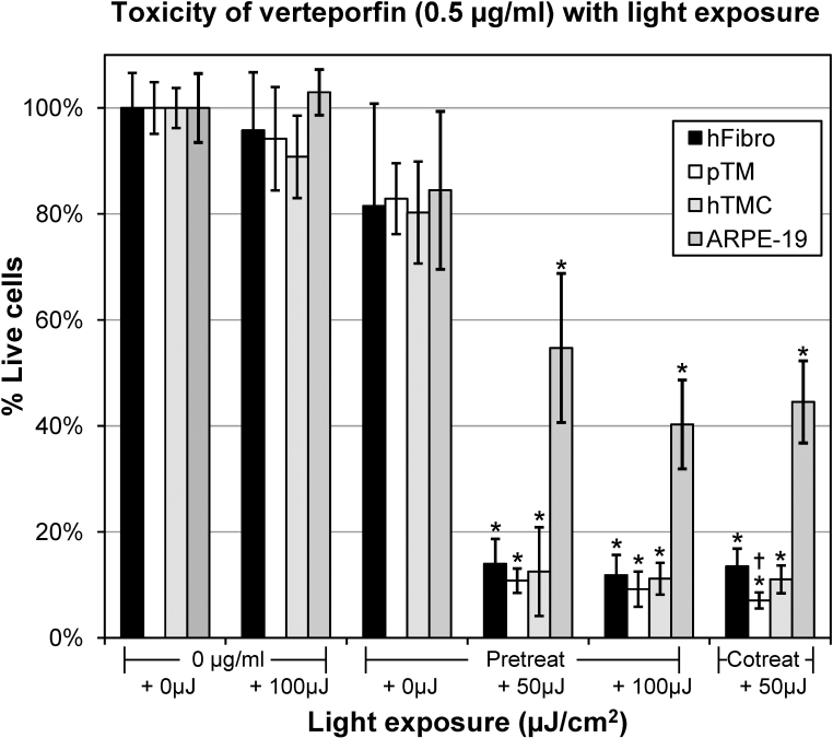
PDT laser treatment alone was insufficient to cause significant cell death in any of the cell types tested. Twenty-four-hour exposure to inactive verteporfin (without PDT laser) caused a dose-dependent decrease in cell viability in hFibro and hTMC, and to a lesser extent ARPE-19 cells. Verteporfin (0.5 µg/ml) without PDT laser activation caused a slight but statistically insignificant reduction in cell viability in hFibro (81.5% ± 19.3%), pTMC (82.9% ± 6.7%), hTMC (80.3% ± 7.7%), and ARPE-19 cells (84.5% ± 14.9%). Verteporfin (0.5 µg/ml) plus 50 µJ/cm(2) PDT laser treatment significantly decreased viability in hFibro (13.5% ± 3.3%), pTMC (7.1% ± 1.5%), hTMC (11.1% ± 5.2%), and ARPE-19 (44.5% ± 7.8%). Similar results were obtained in cells where verteporfin incubation was followed by washout before PDT laser, indicating that verteporfin is internalized by the studied cell lines.
PDT laser-induced cell death was obtained with coincubation of verteporfin or preincubation followed by washout. These results suggest a potential future use of PDT therapy for selective in vivo removal of targeted ocular cells beyond the current use for destroying vascular endothelial cells.
光动力疗法(PDT)激光联合苯并卟啉衍生物维替泊芬是目前治疗脉络膜血管疾病如年龄相关性黄斑变性的临床治疗方法。本研究的目的是检测PDT激光激活和未激活的维替泊芬对各种培养的眼细胞的影响。
用维替泊芬处理原代人巩膜成纤维细胞(hFibro)、原代人小梁网(TM)细胞(hTMC)、原代猪TM细胞(pTMC)和人视网膜色素上皮细胞系(ARPE - 19细胞),其中部分细胞接受PDT激光激活,部分未激活。根据线粒体酶活性(3 -(4,5 - 二甲基 - 2 - 噻唑基)- 2,5 - 二苯基 - 2H - 四氮唑溴盐法)测定细胞活力。
单独的PDT激光治疗不足以在任何测试的细胞类型中引起显著的细胞死亡。未激活的维替泊芬(无PDT激光)24小时暴露导致hFibro和hTMC细胞活力呈剂量依赖性下降,在ARPE - 19细胞中下降程度较小。未经过PDT激光激活的维替泊芬(0.5μg/ml)导致hFibro(81.5%±19.3%)、pTMC(82.9%±6.7%)、hTMC(80.3%±7.7%)和ARPE - 19细胞(84.5%±14.9%)的细胞活力有轻微但无统计学意义的降低。维替泊芬(0.5μg/ml)加50μJ/cm²的PDT激光治疗显著降低了hFibro(13.5%±3.3%)、pTMC(7.1%±1.5%)、hTMC(11.1%±5.2%)和ARPE - 19细胞(44.5%±7.8%)的活力。在维替泊芬孵育后在PDT激光照射前冲洗的细胞中也获得了类似结果,表明维替泊芬被所研究的细胞系内化。
维替泊芬共孵育或预孵育后冲洗可实现PDT激光诱导的细胞死亡。这些结果表明,PDT疗法未来有可能用于在体内选择性去除靶向眼细胞,而不仅仅局限于目前用于破坏血管内皮细胞的用途。