• 文献检索
  • 文档翻译
  • 深度研究
  • 学术资讯
  • Suppr Zotero 插件Zotero 插件
  • 邀请有礼
  • 套餐&价格
  • 历史记录
应用&插件
Suppr Zotero 插件Zotero 插件浏览器插件Mac 客户端Windows 客户端微信小程序
定价
高级版会员购买积分包购买API积分包
服务
文献检索文档翻译深度研究API 文档MCP 服务
关于我们
关于 Suppr公司介绍联系我们用户协议隐私条款
关注我们

Suppr 超能文献

核心技术专利:CN118964589B侵权必究
粤ICP备2023148730 号-1Suppr @ 2026

文献检索

告别复杂PubMed语法,用中文像聊天一样搜索,搜遍4000万医学文献。AI智能推荐,让科研检索更轻松。

立即免费搜索

文件翻译

保留排版,准确专业,支持PDF/Word/PPT等文件格式,支持 12+语言互译。

免费翻译文档

深度研究

AI帮你快速写综述,25分钟生成高质量综述,智能提取关键信息,辅助科研写作。

立即免费体验

血管腔隙内血流斑片状改变时猪冠状动脉管壁 CT 血管造影密度的变化。

Changes in CT angiographic opacification of porcine coronary artery wall with patchy altered flow in vasa vasorum.

机构信息

Department of Physiology and Biomedical Engineering, Rochester, MN 55905, USA.

出版信息

Int J Cardiovasc Imaging. 2013 Aug;29(6):1325-33. doi: 10.1007/s10554-013-0198-6. Epub 2013 Feb 27.

DOI:10.1007/s10554-013-0198-6
PMID:23443339
原文链接:https://pmc.ncbi.nlm.nih.gov/articles/PMC3740031/
Abstract

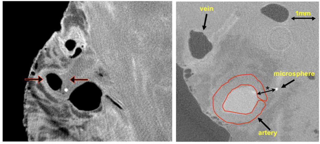
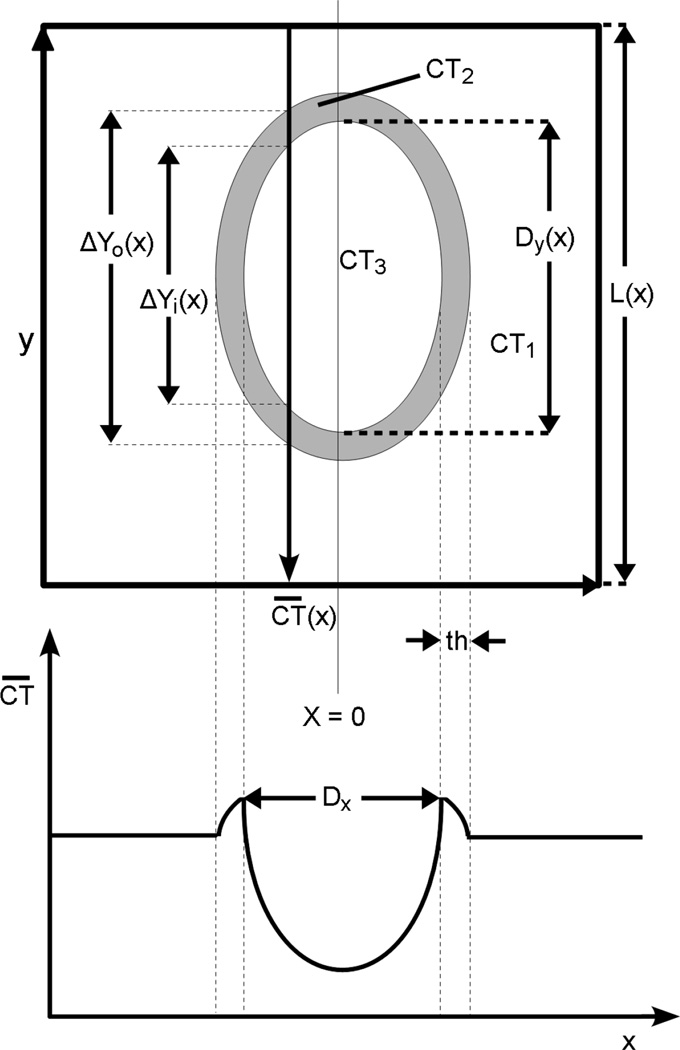
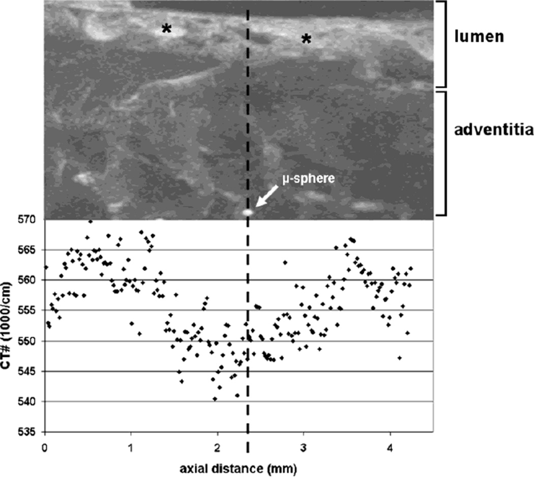
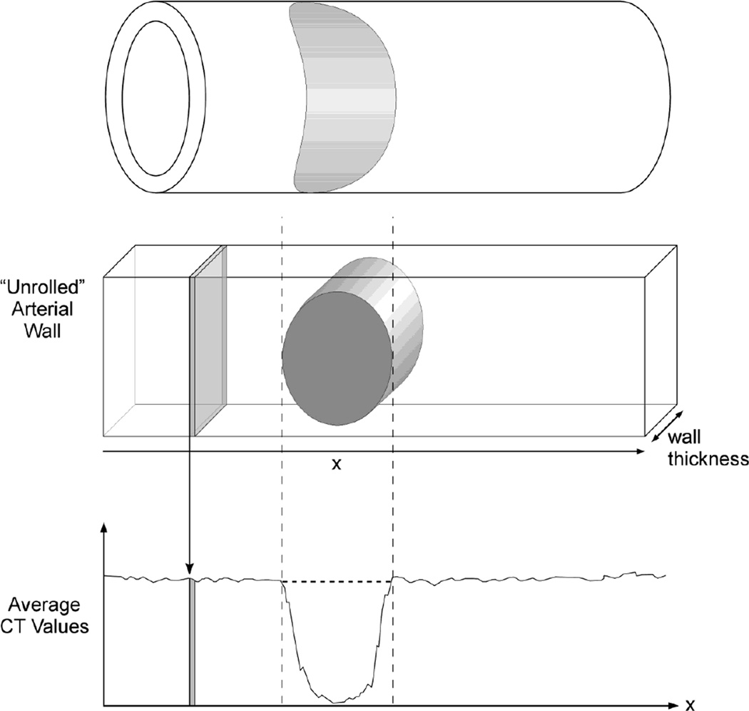
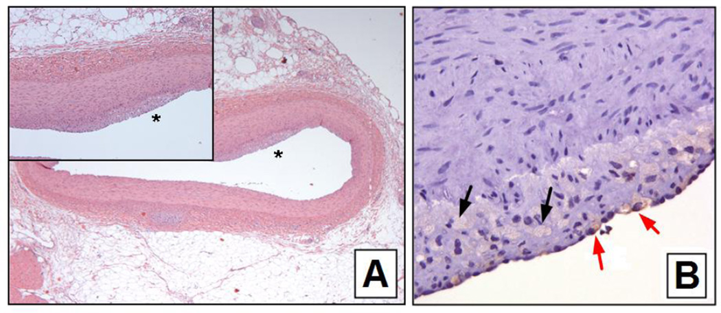
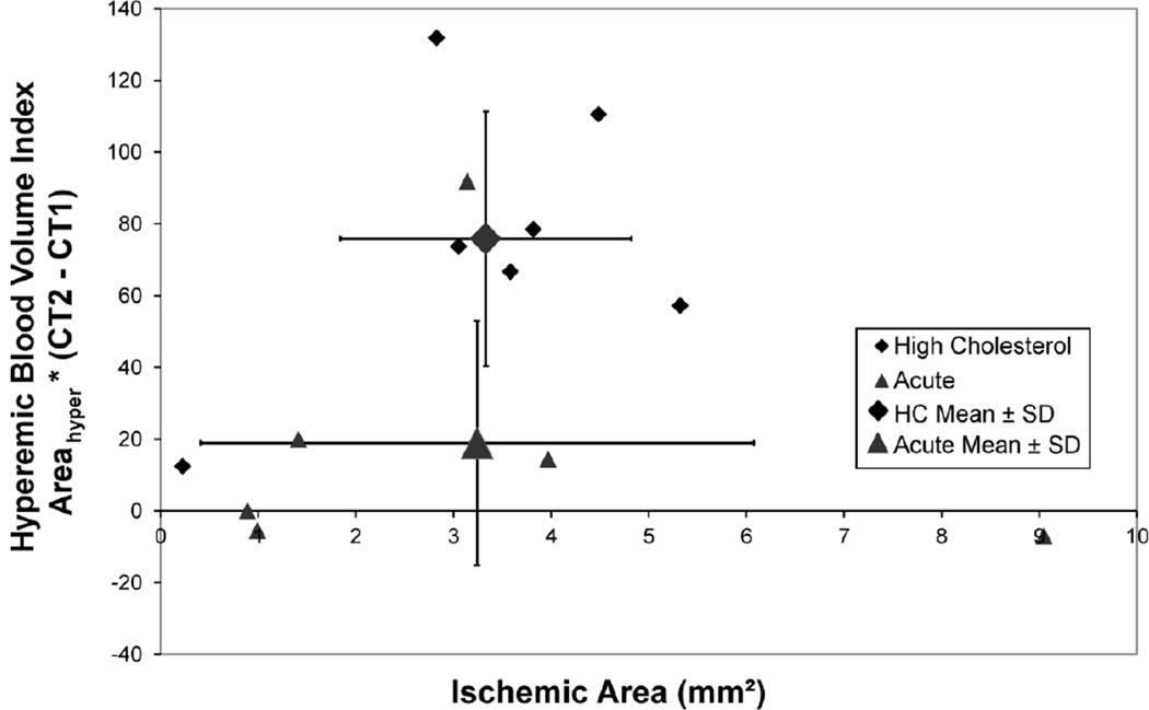
To evaluate the potential of whole-body CT to detect localized areas of decreased or increased vascularity in coronary arterial walls. We used both microsphere embolization of coronary artery vasa vasorum to generate small areas of hypoperfusion and surrounding hyperperfusion of the arterial wall and diet-induced hypercholesterolemia. As a stimulus for localized angiogenesis, such as occurs in early plaque formation in the coronary arterial wall, microspheres were injected selectively into the LAD coronary artery lumens of anesthetized pigs. Fourteen pigs (acute) then had a segment of their LAD harvested during injection of contrast medium and snap-frozen for subsequent cryo-static micro-CT. An additional thirteen pigs (chronic) were allowed to recover, fed a high cholesterol diet and 3 months later were again anesthetized and a segment of the LAD artery harvested and scanned. The spatial distribution of the contrast agent within the arterial wall was measured in contiguous micro-CT images at right angles to the lumen axis with the area of wall in each cross-sectional image being approximately (0.1 mm)(3) in size. In the acute animals there were no localized areas of increased contrast around the hypoperfused embolized perfusion territories in the arterial wall, but in the chronic animals the hypoperfused areas were surrounded by increased contrast. These results suggest that CT might be able to detect localized regions of increased vascularity in the arterial wall as an indicator of early atherosclerotic stimulation of vasa vasorum proliferation.

摘要

为了评估全身 CT 检测冠状动脉壁局部区域血管减少或增加的潜力。我们使用冠状动脉血管丛微球栓塞来产生小面积的灌注不足和动脉壁周围的高灌注,以及饮食诱导的高胆固醇血症。作为局部血管生成的刺激物,如在冠状动脉壁早期斑块形成中发生的那样,微球被选择性地注入麻醉猪的 LAD 冠状动脉管腔中。然后,14 只猪(急性)在注射对比剂时采集其 LAD 的一段,并立即冷冻用于随后的冷冻静态 micro-CT。另外 13 只猪(慢性)被允许恢复,给予高胆固醇饮食,3 个月后再次麻醉并采集一段 LAD 动脉进行扫描。在与管腔轴成直角的连续 micro-CT 图像中测量动脉壁内造影剂的空间分布,每个横截面图像中的壁面积约为 (0.1 mm)(3)。在急性动物中,在灌注不足的栓塞灌注区域周围没有局部区域的对比度增加,但在慢性动物中,灌注不足区域被对比度增加所包围。这些结果表明,CT 可能能够检测到动脉壁中局部区域的血管增多,作为血管丛增殖的早期动脉粥样硬化刺激的指标。

https://cdn.ncbi.nlm.nih.gov/pmc/blobs/1c8b/3740031/9132f98731f2/nihms450076f7.jpg
https://cdn.ncbi.nlm.nih.gov/pmc/blobs/1c8b/3740031/f8ca979c838b/nihms450076f1.jpg
https://cdn.ncbi.nlm.nih.gov/pmc/blobs/1c8b/3740031/b343c3017cc0/nihms450076f2.jpg
https://cdn.ncbi.nlm.nih.gov/pmc/blobs/1c8b/3740031/af48066c0cb4/nihms450076f3.jpg
https://cdn.ncbi.nlm.nih.gov/pmc/blobs/1c8b/3740031/78b24d8135df/nihms450076f4.jpg
https://cdn.ncbi.nlm.nih.gov/pmc/blobs/1c8b/3740031/1b65f585bad5/nihms450076f5.jpg
https://cdn.ncbi.nlm.nih.gov/pmc/blobs/1c8b/3740031/ff7238723b95/nihms450076f6.jpg
https://cdn.ncbi.nlm.nih.gov/pmc/blobs/1c8b/3740031/9132f98731f2/nihms450076f7.jpg

相似文献

1
Changes in CT angiographic opacification of porcine coronary artery wall with patchy altered flow in vasa vasorum.血管腔隙内血流斑片状改变时猪冠状动脉管壁 CT 血管造影密度的变化。
Int J Cardiovasc Imaging. 2013 Aug;29(6):1325-33. doi: 10.1007/s10554-013-0198-6. Epub 2013 Feb 27.
2
IVUS detection of vasa vasorum blood flow distribution in coronary artery vessel wall.血管内超声检测冠状动脉血管壁中血管的血流分布。
JACC Cardiovasc Imaging. 2012 Sep;5(9):935-40. doi: 10.1016/j.jcmg.2011.12.027.
3
Quantification of vasa vasorum density in multi-slice computed tomographic coronary angiograms: role of computed tomographic image voxel size.多层螺旋计算机断层扫描冠状动脉造影中血管滋养管密度的定量分析:计算机断层扫描图像体素大小的作用
J Comput Assist Tomogr. 2010 Mar-Apr;34(2):273-8. doi: 10.1097/RCT.0b013e3181bb0d32.
4
Role of vasa vasorum in transendothelial solute transport in the coronary vessel wall: a study with cryostatic micro-CT.血管滋养血管在冠状动脉壁内皮溶质转运中的作用:一项低温微型计算机断层扫描研究
Am J Physiol Heart Circ Physiol. 2004 Nov;287(5):H2346-51. doi: 10.1152/ajpheart.00066.2004. Epub 2004 Jun 3.
5
Impact of coronary vasa vasorum functional structure on coronary vessel wall perfusion distribution.冠状动脉滋养血管功能结构对冠状动脉壁灌注分布的影响。
Am J Physiol Heart Circ Physiol. 2003 Nov;285(5):H2019-26. doi: 10.1152/ajpheart.00399.2003. Epub 2003 Jul 10.
6
Increased spatial vasa vasorum density in the proximal LAD in hypercholesterolemia--implications for vulnerable plaque-development.高胆固醇血症患者左前降支近端血管滋养血管空间密度增加——对易损斑块形成的影响
Atherosclerosis. 2007 Jun;192(2):246-52. doi: 10.1016/j.atherosclerosis.2006.07.004. Epub 2006 Aug 21.
7
Enhanced coronary vasa vasorum neovascularization in experimental hypercholesterolemia.实验性高胆固醇血症中冠状动脉血管滋养血管新生血管形成增强。
J Clin Invest. 1998 Apr 15;101(8):1551-6. doi: 10.1172/JCI1568.
8
Chronic endothelin receptor antagonism prevents coronary vasa vasorum neovascularization in experimental hypercholesterolemia.慢性内皮素受体拮抗作用可预防实验性高胆固醇血症中冠状动脉血管滋养管新生血管形成。
J Am Coll Cardiol. 2002 May 1;39(9):1555-61. doi: 10.1016/s0735-1097(02)01798-9.
9
Functional anatomy and hemodynamic characteristics of vasa vasorum in the walls of porcine coronary arteries.猪冠状动脉壁内血管滋养血管的功能解剖和血流动力学特征。
Anat Rec A Discov Mol Cell Evol Biol. 2003 Jun;272(2):526-37. doi: 10.1002/ar.a.10060.
10
Experimental hypercholesterolemia differentially affects adventitial vasa vasorum and vessel structure of the left internal thoracic and coronary arteries.实验性高胆固醇血症对左胸廓内动脉和冠状动脉的外膜血管滋养管及血管结构有不同影响。
J Thorac Cardiovasc Surg. 2005 Apr;129(4):767-72. doi: 10.1016/j.jtcvs.2004.08.014.

引用本文的文献

1
Atherosclerosis and atheroma plaque rupture: imaging modalities in the visualization of vasa vasorum and atherosclerotic plaques.动脉粥样硬化与动脉粥样斑块破裂:滋养血管和动脉粥样硬化斑块可视化中的成像方式
ScientificWorldJournal. 2014 Feb 11;2014:312764. doi: 10.1155/2014/312764. eCollection 2014.
2
Cardiovascular imaging 2013 in the International Journal of Cardiovascular Imaging.《国际心血管影像杂志》2013年心血管成像相关内容
Int J Cardiovasc Imaging. 2014 Apr;30(4):683-95. doi: 10.1007/s10554-014-0405-0.

本文引用的文献

1
Computed tomography coronary angiography in asymptomatic patients.计算机断层扫描冠状动脉造影在无症状患者中的应用。
Radiol Med. 2011 Dec;116(8):1161-73. doi: 10.1007/s11547-011-0723-4. Epub 2011 Sep 2.
2
Least squares parameter estimation methods for material decomposition with energy discriminating detectors.基于能量甄别探测器的物质分解的最小二乘参数估计方法。
Med Phys. 2011 Jan;38(1):245-55. doi: 10.1118/1.3525840.
3
Positive remodeling of the coronary arteries detected by magnetic resonance imaging in an asymptomatic population: MESA (Multi-Ethnic Study of Atherosclerosis).
磁共振成像检测到无症状人群冠状动脉的正向重塑:动脉粥样硬化多族裔研究(MESA)
J Am Coll Cardiol. 2009 May 5;53(18):1708-15. doi: 10.1016/j.jacc.2008.12.063.
4
Role of vasa vasorum in transendothelial solute transport in the coronary vessel wall: a study with cryostatic micro-CT.血管滋养血管在冠状动脉壁内皮溶质转运中的作用:一项低温微型计算机断层扫描研究
Am J Physiol Heart Circ Physiol. 2004 Nov;287(5):H2346-51. doi: 10.1152/ajpheart.00066.2004. Epub 2004 Jun 3.
5
Accuracy of multidetector spiral computed tomography in identifying and differentiating the composition of coronary atherosclerotic plaques: a comparative study with intracoronary ultrasound.多排螺旋计算机断层扫描在识别和区分冠状动脉粥样硬化斑块成分方面的准确性:与冠状动脉内超声的比较研究
J Am Coll Cardiol. 2004 Apr 7;43(7):1241-7. doi: 10.1016/j.jacc.2003.10.059.
6
Detection of calcified and noncalcified coronary atherosclerotic plaque by contrast-enhanced, submillimeter multidetector spiral computed tomography: a segment-based comparison with intravascular ultrasound.通过对比增强亚毫米多层螺旋计算机断层扫描检测钙化和非钙化冠状动脉粥样硬化斑块:与血管内超声的节段性比较
Circulation. 2004 Jan 6;109(1):14-7. doi: 10.1161/01.CIR.0000111517.69230.0F. Epub 2003 Dec 22.
7
Non-invasive assessment of plaque morphology and remodeling in mildly stenotic coronary segments: comparison of 16-slice computed tomography and intravascular ultrasound.轻度狭窄冠状动脉节段斑块形态和重构的无创评估:16层计算机断层扫描与血管内超声的比较
Coron Artery Dis. 2003 Sep;14(6):459-62. doi: 10.1097/00019501-200309000-00007.
8
Impact of coronary vasa vasorum functional structure on coronary vessel wall perfusion distribution.冠状动脉滋养血管功能结构对冠状动脉壁灌注分布的影响。
Am J Physiol Heart Circ Physiol. 2003 Nov;285(5):H2019-26. doi: 10.1152/ajpheart.00399.2003. Epub 2003 Jul 10.
9
Computed tomography and magnetic resonance imaging for noninvasive coronary angiography and plaque imaging: current and potential future concepts.用于无创冠状动脉造影和斑块成像的计算机断层扫描和磁共振成像:当前及未来潜在概念
Circulation. 2002 Oct 8;106(15):2026-34. doi: 10.1161/01.cir.0000034392.34211.fc.
10
Cryostatic micro-computed tomography imaging of arterial wall perfusion.动脉壁灌注的低温显微计算机断层扫描成像
Scanning. 2002 Jul-Aug;24(4):186-90. doi: 10.1002/sca.4950240405.