Commonwealth Scientific and Industrial Research Organisation (CSIRO), Perth, WA, Australia.
Transl Psychiatry. 2013 Feb 26;3(2):e233. doi: 10.1038/tp.2012.150.
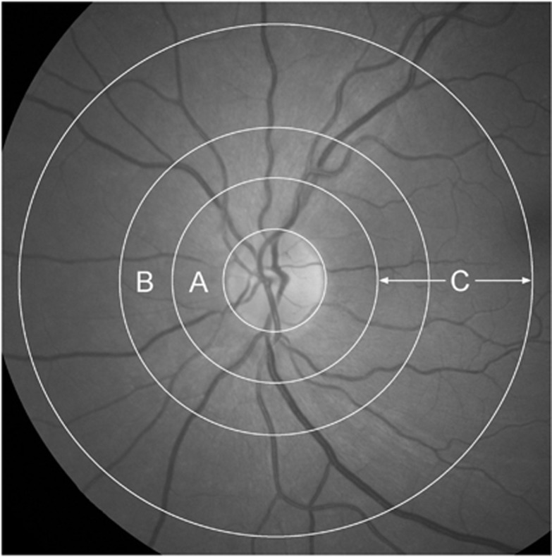
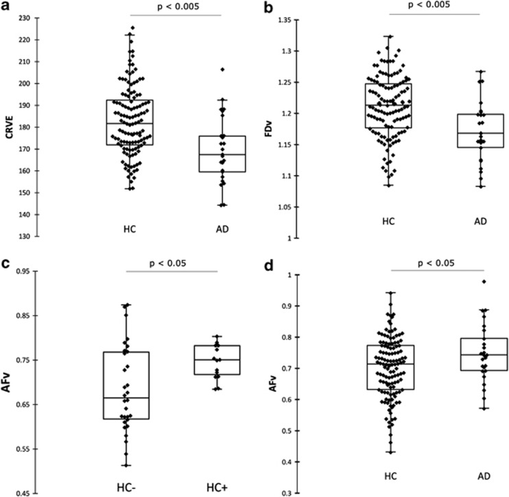
The earliest detectable change in Alzheimer's disease (AD) is the buildup of amyloid plaque in the brain. Early detection of AD, prior to irreversible neurological damage, is important for the efficacy of current interventions as well as for the development of new treatments. Although PiB-PET imaging and CSF amyloid are the gold standards for early AD diagnosis, there are practical limitations for population screening. AD-related pathology occurs primarily in the brain, but some of the hallmarks of the disease have also been shown to occur in other tissues, including the retina, which is more accessible for imaging. Retinal vascular changes and degeneration have previously been reported in AD using optical coherence tomography and laser Doppler techniques. This report presents results from analysis of retinal photographs from AD and healthy control participants from the Australian Imaging, Biomarkers and Lifestyle (AIBL) Flagship Study of Ageing. This is the first study to investigate retinal blood vessel changes with respect to amyloid plaque burden in the brain. We demonstrate relationships between retinal vascular parameters, neocortical brain amyloid plaque burden and AD. A number of RVPs were found to be different in AD. Two of these RVPs, venular branching asymmetry factor and arteriolar length-to-diameter ratio, were also higher in healthy individuals with high plaque burden (P = 0.01 and P = 0.02 respectively, after false discovery rate adjustment). Retinal photographic analysis shows potential as an adjunct for early detection of AD or monitoring of AD-progression or response to treatments.
阿尔茨海默病(AD)最早可检测到的变化是大脑中淀粉样斑块的堆积。在不可逆的神经损伤之前,早期发现 AD 对于当前干预措施的疗效以及新疗法的开发都很重要。虽然 PiB-PET 成像和 CSF 淀粉样蛋白是 AD 早期诊断的金标准,但在人群筛查方面存在实际限制。AD 相关的病理学主要发生在大脑中,但该疾病的一些特征也已在其他组织中显示出来,包括视网膜,视网膜更适合进行成像。先前已经使用光学相干断层扫描和激光多普勒技术在 AD 中报告了视网膜血管变化和退行性变。本报告介绍了来自澳大利亚成像、生物标志物和生活方式(AIBL)老龄化旗舰研究的 AD 和健康对照参与者的视网膜照片分析结果。这是第一项研究,旨在调查与大脑中淀粉样斑块负担有关的视网膜血管变化。我们证明了视网膜血管参数、大脑新皮质淀粉样斑块负担和 AD 之间存在关系。AD 患者存在多种 RVPs 差异。其中两种 RVPs,静脉分支不对称因子和小动脉长度与直径比,在斑块负担高的健康个体中也更高(经假发现率调整后分别为 P = 0.01 和 P = 0.02)。视网膜摄影分析显示出作为 AD 早期检测或 AD 进展或治疗反应监测的辅助手段的潜力。