Neuroradiology Division, Massachusetts General Hospital, Harvard Medical School, Boston, Massachusetts 02114, USA.
J Neurointerv Surg. 2013 May;5 Suppl 1(Suppl 1):i7-12. doi: 10.1136/neurintsurg-2013-010715. Epub 2013 Mar 14.
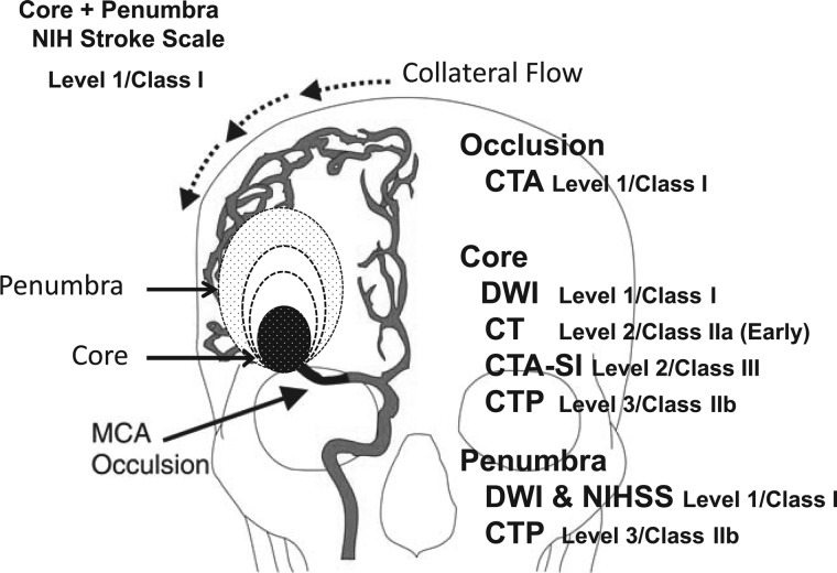
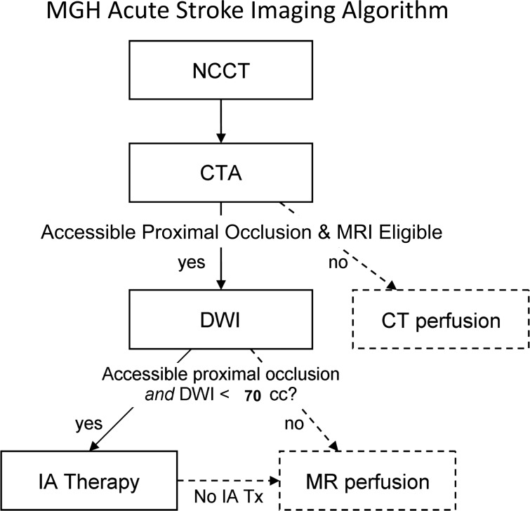
The Massachusetts General Hospital Neuroradiology Division employed an experience and evidence based approach to develop a neuroimaging algorithm to best select patients with severe ischemic strokes caused by anterior circulation occlusions (ACOs) for intravenous tissue plasminogen activator and endovascular treatment. Methods found to be of value included the National Institutes of Health Stroke Scale (NIHSS), non-contrast CT, CT angiography (CTA) and diffusion MRI. Perfusion imaging by CT and MRI were found to be unnecessary for safe and effective triage of patients with severe ACOs. An algorithm was adopted that includes: non-contrast CT to identify hemorrhage and large hypodensity followed by CTA to identify the ACO; diffusion MRI to estimate the core infarct; and NIHSS in conjunction with diffusion data to estimate the clinical penumbra.
马萨诸塞州综合医院神经放射科采用了一种基于经验和证据的方法来制定神经影像学算法,以最佳选择因前循环闭塞(ACO)导致的严重缺血性中风患者进行静脉组织型纤溶酶原激活剂和血管内治疗。被发现有价值的方法包括国立卫生研究院中风量表(NIHSS)、非对比 CT、CT 血管造影(CTA)和弥散 MRI。发现 CT 和 MRI 灌注成像对于严重 ACO 患者的安全有效分诊是不必要的。采用了一种算法,包括:非对比 CT 以识别出血和大低密影,然后进行 CTA 以识别 ACO;弥散 MRI 以估计核心梗死;以及 NIHSS 与弥散数据结合以估计临床半影区。