Department of Anatomy and Neurobiology, Northeast Ohio Medical University Rootstown, OH, USA.
Front Neural Circuits. 2013 Mar 20;7:41. doi: 10.3389/fncir.2013.00041. eCollection 2013.
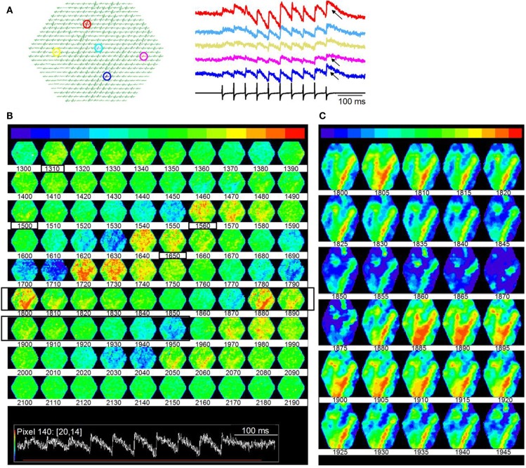
We used optical imaging with voltage-sensitive dyes to investigate the spatio-temporal dynamics of synaptically evoked activity in brain slices of the inferior colliculus (IC). Responses in transverse slices which preserve cross-frequency connections and in modified sagittal slices that preserve connections within frequency laminae were evoked by activating the lateral lemniscal tract. Comparing activity between small and large populations of cells revealed response areas in the central nucleus of the IC that were similar in magnitude but graded temporally. In transverse sections, these response areas are summed to generate a topographic response profile. Activity through the commissure to the contralateral IC required an excitation threshold that was reached when GABAergic inhibition was blocked. Within laminae, module interaction created temporal homeostasis. Diffuse activity evoked by a single lemniscal shock re-organized into distinct spatial and temporal compartments when stimulus trains were used, and generated a directional activity profile within the lamina. Using different stimulus patterns to activate subsets of microcircuits in the central nucleus of the IC, we found that localized responses evoked by low-frequency stimulus trains spread extensively when train frequency was increased, suggesting recruitment of silent microcircuits. Long stimulus trains activated a circuit specific to post-inhibitory rebound neurons. Rebound microcircuits were defined by a focal point of initiation that spread to an annular ring that oscillated between inhibition and excitation. We propose that much of the computing power of the IC is derived from local circuits, some of which are cell-type specific. These circuits organize activity within and across frequency laminae, and are critical in determining the stimulus-selectivity of auditory coding.
我们使用电压敏感染料的光学成像技术,研究了下丘脑中脑切片(IC)中突触诱发活动的时空动态。通过激活外侧丘系来诱发横向切片(保留了交叉频率连接)和改良矢状切片(保留了频率层内的连接)中的反应。比较小细胞和大细胞群体的活动,揭示了 IC 中央核中的反应区,其幅度相似,但时间上呈梯度变化。在横断面上,这些反应区被叠加以生成地形反应轮廓。通过连合向对侧 IC 的活动需要达到 GABA 能抑制被阻断时的激发阈值。在层内,模块相互作用产生了时间内稳态。当使用刺激序列时,单个丘系冲击引起的弥散活动重新组织成不同的空间和时间区室,并在层内产生了定向活动轮廓。使用不同的刺激模式来激活 IC 中央核中的微电路子集,我们发现低频刺激序列引起的局部反应在增加刺激序列频率时会广泛扩散,这表明沉默微电路的招募。长刺激序列激活了特定于抑制后反弹神经元的回路。反弹微电路的特征是起始焦点,其扩散到一个环形环,该环形环在抑制和兴奋之间振荡。我们提出,IC 的大部分计算能力来自于局部回路,其中一些回路是细胞类型特异性的。这些回路组织了层内和层间的活动,对听觉编码的刺激选择性至关重要。