Maeda Masashi, Kasahara Naoya, Doi Junshi, Iijima Yuki, Kikuchi Takeshi, Teratani Takumi, Kobayashi Eiji
Division of Development of Advanced Therapy , Center for Development of Advanced Medical Technology, Jichi Medical University , Tochigi , Japan.
Heart Asia. 2013 Jan 17;5(1):7-14. doi: 10.1136/heartasia-2012-010160. Print 2013.
We developed a novel luciferase-based viability assay for assessing the viability of hearts preserved in different solutions. We examined whether this system could predict heart damage and survival after transplantation in rats.
By our novel system, preserved heart viability evaluation and transplanted heart-graft functional research study.
University basic science laboratory.
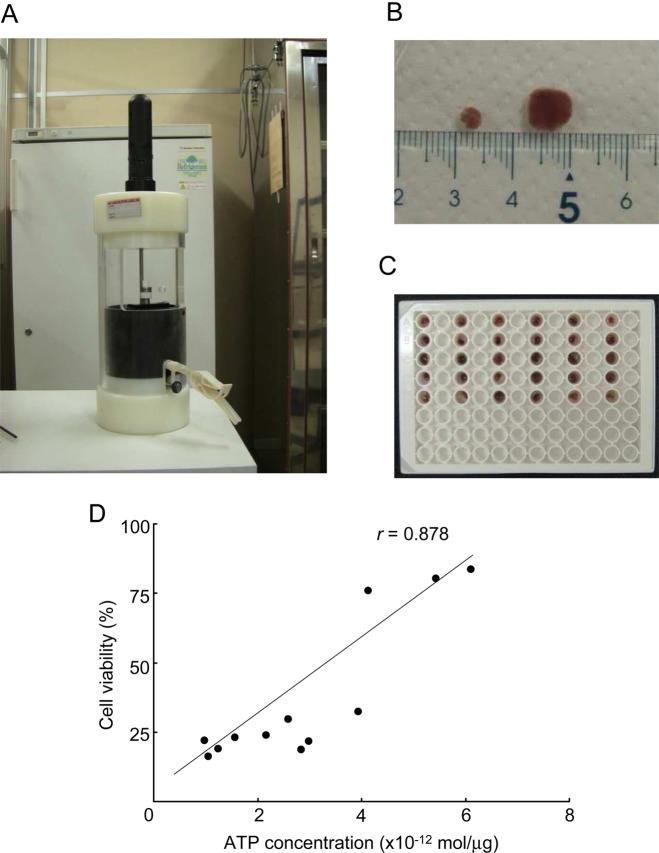
Isolated Luciferase-transgenic Lewis (LEW) rat cardiac-tissue-chips were plated on 96-well tissue-culture plates and incubated in preservation solutions at 4°C. Viability was measured as photon intensity by using a bio-imaging system. Heart-grafts preserved in University of Wisconsin (UW), extracellular-trehalose-Kyoto (ETK), Euro-Collins (EC), histidin-tryptophan-ketoglutarat solution (HTK), lactated Ringer's (LR) or normal saline solution were transplanted cervically by using a cuff-technique or into the abdomens of syngeneic wild-type LEW rats by using conventional microsurgical suture techniques.
Imaging an evaluation of preservation heart-graft and functional analysis.
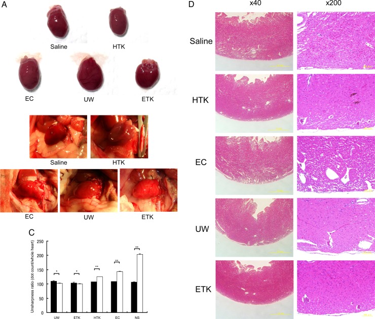
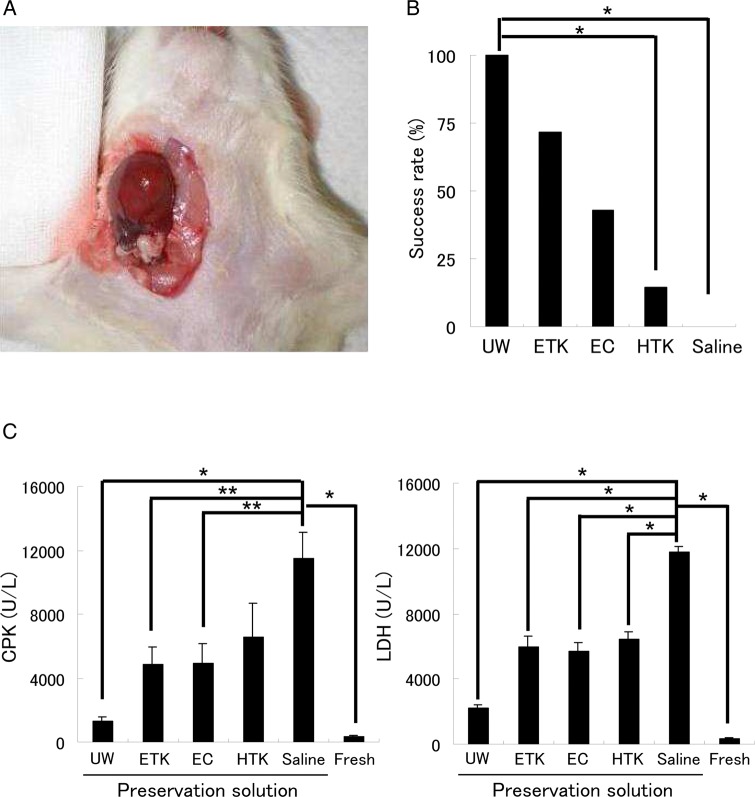
Cardiac-tissue-chips preserved with UW, HTK or ETK solution gave higher luminance than those preserved with EC, LR or normal saline (p<0.03). After 24 h of preservation of hearts in each solution at 4°C, the beating of the isolated hearts was evaluated. The success rate, evaluation of beating, of cervical heart transplants using UW and ETK solution exceeded 70%, but those using other preservation solutions were lower (UW: 100%, ETK: 75%, EC: 42.86%, HTK: 14.29%, normal saline: 0%). Histological analysis of cervical heart-grafts after 3 h preservation by myeloperoxidase (MPO), zona occludens-1(ZO-1), and caspase-3 immunostaining revealed different degrees of preservation damage in all grafts.
Our novel assay system is simple and can test multiple solutions. It should therefore be a powerful tool for developing and improving new heart-graft preservation solutions.
我们开发了一种基于荧光素酶的新型活力测定法,用于评估保存在不同溶液中的心脏的活力。我们研究了该系统是否能够预测大鼠心脏移植后的损伤和存活情况。
通过我们的新型系统进行保存心脏活力评估和移植心脏移植物功能研究。
大学基础科学实验室。
将分离的荧光素酶转基因Lewis(LEW)大鼠心脏组织芯片接种到96孔组织培养板上,并于4°C在保存溶液中孵育。使用生物成像系统将活力测量为光子强度。使用袖带技术将保存在威斯康星大学(UW)溶液、细胞外海藻糖-京都(ETK)溶液、欧洲柯林斯(EC)溶液、组氨酸-色氨酸-酮戊二酸溶液(HTK)、乳酸林格氏液(LR)或生理盐水中的心脏移植物移植到同基因野生型LEW大鼠的颈部,或使用传统显微外科缝合技术将其移植到腹部。
成像评估保存心脏移植物并进行功能分析。
用UW、HTK或ETK溶液保存的心脏组织芯片比用EC、LR或生理盐水保存的芯片具有更高的亮度(p<0.03)。在4°C下将心脏在每种溶液中保存24小时后,评估离体心脏的跳动情况。使用UW和ETK溶液进行颈部心脏移植的成功率(跳动评估)超过70%,但使用其他保存溶液的成功率较低(UW:100%,ETK:75%,EC:42.86%,HTK:14.29%,生理盐水:0%)。通过髓过氧化物酶(MPO)、紧密连接蛋白-1(ZO-1)和半胱天冬酶-3免疫染色对保存3小时后的颈部心脏移植物进行组织学分析,结果显示所有移植物均有不同程度的保存损伤。
我们的新型检测系统简单且可测试多种溶液。因此,它应该是开发和改进新型心脏移植物保存溶液的有力工具。