Department of Oral and Maxillofacial Surgery, Hanover Medical School, Carl-Neuberg-Strasse 1, Hanover D-30625, Germany.
Head Face Med. 2013 Apr 20;9:14. doi: 10.1186/1746-160X-9-14.
To compare two methods of creating three-dimensional representations of mandibular cysts and tumors on the basis of computed tomography (CT) and cone beam computed tomography (CBCT) data.
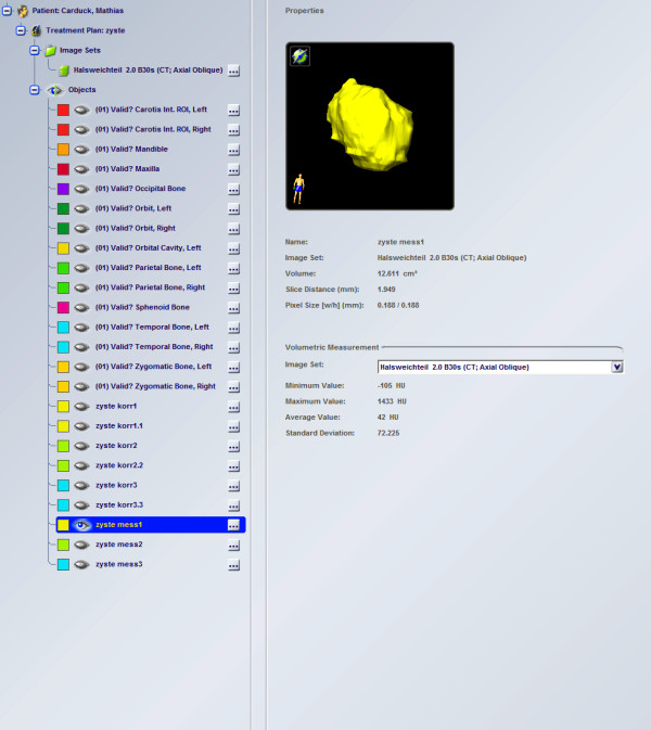
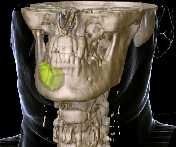
A total of 71 patients with acquired jaw cysts took part in this retrospective clinical study. CT and CBCT scans were obtained from all patients and saved in the Digital Imaging and Communications in Medicine (DICOM) format. Data were analyzed twice with iPlan software. Analysis was performed manually and using an interpolarization algorithm. The accuracy of the two methods in assessing cyst volume was compared.
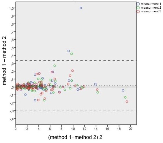
Manual delineation did not provide more accurate results than the interpolarization algorithm.
There are no major differences between manual analysis and analysis using the interpolarization algorithm. The use of the algorithm, however, has the advantage of rapidity.
基于 CT 和锥形束 CT 数据,比较两种生成下颌骨囊肿和肿瘤三维图像的方法。
本回顾性临床研究共纳入 71 例获得性颌骨囊肿患者。对所有患者进行 CT 和 CBCT 扫描,并以 DICOM 格式保存。数据使用 iPlan 软件进行了两次分析。分析分别手动进行和使用极间算法进行。比较了两种方法评估囊肿体积的准确性。
手动勾画并未比极间算法提供更准确的结果。
手动分析与使用极间算法分析之间没有明显差异。然而,算法的使用具有快速的优点。