Center for Psychiatric Research, Department of Clinical Neuroscience, Karolinska Institutet, Stockholm SE-171 77, Sweden.
EJNMMI Res. 2013 Apr 24;3(1):30. doi: 10.1186/2191-219X-3-30.
The activation of microglia, in general, and the upregulation of the translocator protein (18 kDa) (TSPO) system, in particular, are key features of neuroinflammation, of which the in vivo visualization and quantitative assessment are still challenging due to the lack of appropriate molecular imaging biomarkers. Recent positron emission tomography (PET) studies using TSPO radioligands such as [11C]PK11195 and [11C]PBR28 have indicated the usefulness of these PET biomarkers in patients with neuroinflammatory diseases, including multiple sclerosis (MS). [18F]FEDAA1106 is a recently developed PET radioligand for the in vivo quantification of TSPO. In the present study, we aimed at investigating the diagnostic usefulness of [18F]FEDAA1106 in patients with MS.
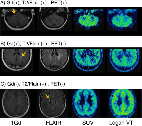
Nine patients (three on the interferon beta therapy and six without immunomodulatory therapy; seven females/two males; age 34.2 ± 9.1 years old) with relapsing-remitting MS in acute relapse and with gadolinium (Gd)-enhancing lesion(s) in the magnetic resonance imaging (MRI) scans and five healthy controls (four females/one male, age 38.0 ± 9.7 years old) were investigated in this study. Genetic information about the TSPO binding could not be obtained because knowledge about the importance of genetic background for TSPO binding was not available at the time the study was performed. Dynamic PET measurements were performed using an ECAT EXACT HR system (CTI/Siemens, Knoxville, TN, USA) for a total of 150 min, with a 30-min break after the injection of 153.4 ± 10.2 MBq of [18F]FEDAA1106. Metabolite-corrected arterial plasma samples were used to calculate the input function. PET data were analyzed in the following ways: (1) region-of-interest analysis for cortical and subcortical regions was performed using a two-tissue compartment kinetic model in order to estimate binding potentials (BPND) and distribution volume (VT), (2) the feasibility of the estimation of BPND and VT was investigated for MS lesions, and (3) VT parametric images by a Logan plot and standard uptake value (SUV) images were visually compared with the corresponding MRI, focusing on MRI-identified MS lesions.
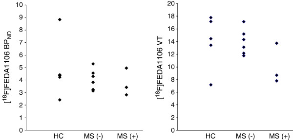
There were no significant differences in the BPND or VT values between patients with MS and healthy controls. Robust BPND and VT values could not be obtained for most MS lesions due to noisy time-activity curves. Visual inspection of VT and SUV images in all nine patients did not reveal high uptake of the radioligand inside and beyond MRI-identified active MS lesions with the exception of one Gd-enhanced MS lesion in the whole patient population.
In our study, [18F]FEDAA1106 as a PET radioligand could neither differentiate patients with MS from healthy controls nor detect active plaques in the brain of MS patients. Stratification with respect to genetics and binder status might help to uncover the differences between the groups, which could not be detected here.
ClinicalTrials.gov, NCT01031199.
小胶质细胞的激活,一般来说,以及转位蛋白(18 kDa)(TSPO)系统的上调,是神经炎症的关键特征,由于缺乏适当的分子影像学生物标志物,其体内可视化和定量评估仍然具有挑战性。最近使用 TSPO 放射性配体(如[11C]PK11195和[11C]PBR28)的正电子发射断层扫描(PET)研究表明,这些 PET 生物标志物在神经炎症性疾病患者中具有一定的应用价值,包括多发性硬化症(MS)。[18F]FEDAA1106 是一种最近开发的用于体内定量 TSPO 的 PET 放射性配体。在本研究中,我们旨在研究[18F]FEDAA1106 在 MS 患者中的诊断价值。
本研究纳入了 9 名处于急性复发期的复发缓解型 MS 患者(3 名接受干扰素β治疗,6 名未接受免疫调节治疗;7 名女性/2 名男性;年龄 34.2±9.1 岁)和 5 名健康对照者(4 名女性/1 名男性;年龄 38.0±9.7 岁)。由于当时还没有关于 TSPO 结合遗传背景重要性的知识,因此无法获得有关 TSPO 结合的遗传信息。使用 ECAT EXACT HR 系统(CTI/Siemens,田纳西州诺克斯维尔)进行了 150 分钟的动态 PET 测量,在注射 153.4±10.2MBq [18F]FEDAA1106 后进行了 30 分钟的休息。使用代谢校正的动脉血浆样本计算输入函数。以以下方式分析 PET 数据:(1)使用双组织室动力学模型进行皮质和皮质下区域的感兴趣区分析,以估计结合潜能(BPND)和分布容积(VT);(2)研究 MS 病变中 BPND 和 VT 估计的可行性;(3)通过 Logan 图和标准摄取值(SUV)图像进行 VT 参数图像的视觉比较,并重点关注 MRI 确定的 MS 病变。
MS 患者和健康对照者的 BPND 或 VT 值无显著差异。由于时间活性曲线噪声较大,大多数 MS 病变的 BPND 和 VT 值均无法获得。对所有 9 名患者的 VT 和 SUV 图像进行视觉检查,除了整个患者群体中的一个 Gd 增强 MS 病变外,并未发现放射性配体在 MRI 确定的活跃 MS 病变内和病变外的摄取增加。
在我们的研究中,[18F]FEDAA1106 作为一种 PET 放射性配体,既不能区分 MS 患者与健康对照者,也不能检测 MS 患者大脑中的活动斑块。对遗传学和配体状态进行分层可能有助于揭示两组之间的差异,而这在本研究中无法检测到。
ClinicalTrials.gov,NCT01031199。