Department of Medicine, Division of Genomic Medicine, George Washington University School of Medicine and Health Sciences, Washington DC, USA.
Int J Biol Sci. 2013 Apr 22;9(4):350-60. doi: 10.7150/ijbs.6058. Print 2013.
Anthracyclines, such as doxorubicin (Adriamycin), are highly effective chemotherapeutic agents, but are well known to cause myocardial dysfunction and life-threatening congestive heart failure (CHF) in some patients.
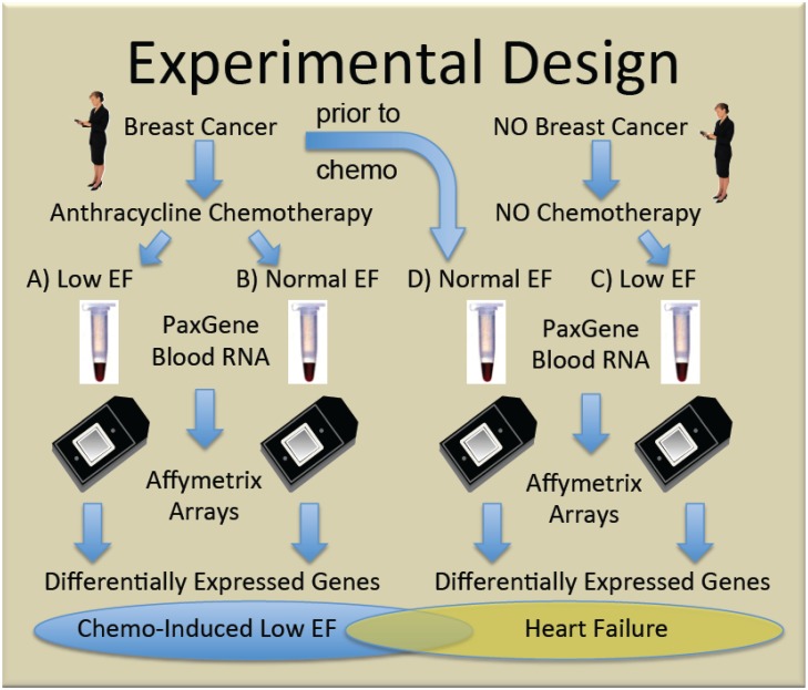
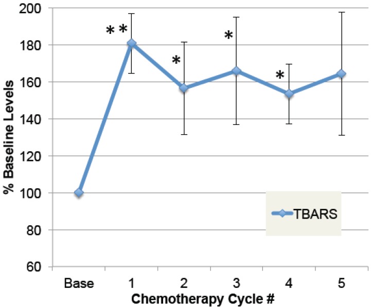
To generate new hypotheses about its etiology, genome-wide transcript analysis was performed on whole blood RNA from women that received doxorubicin-based chemotherapy and either did, or did not develop CHF, as defined by ejection fractions (EF)≤40%. Women with non-ischemic cardiomyopathy unrelated to chemotherapy were compared to breast cancer patients prior to chemo with normal EF to identify heart failure-related transcripts in women not receiving chemotherapy. Byproducts of oxidative stress in plasma were measured in a subset of patients.
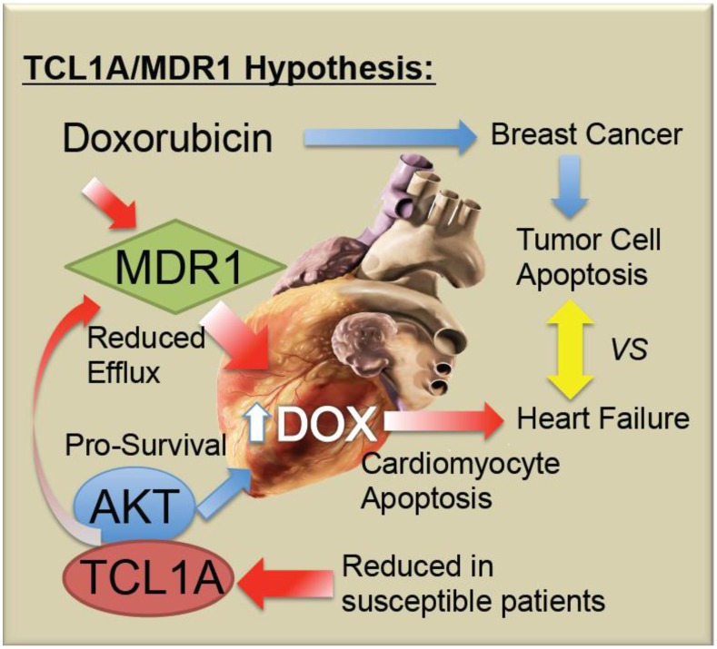
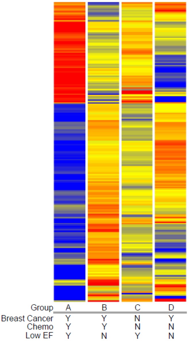
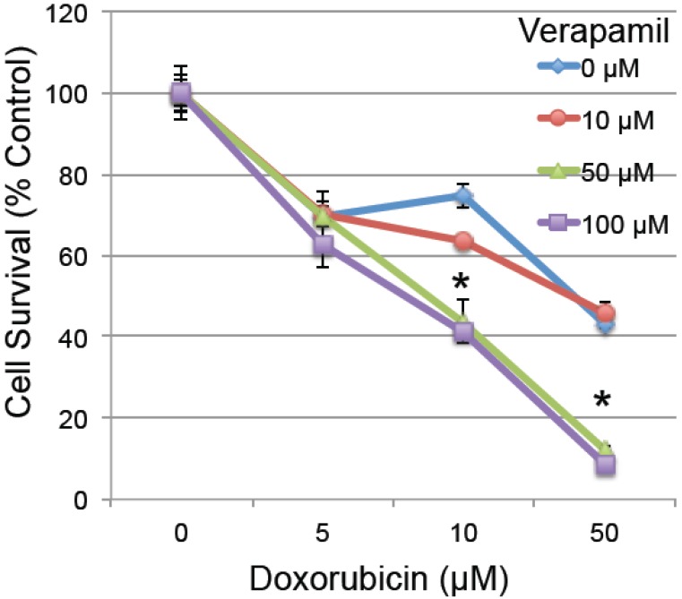
The results indicate that patients treated with doxorubicin showed sustained elevations in oxidative byproducts in plasma. At the RNA level, women who exhibited low EFs after chemotherapy had 260 transcripts that differed >2-fold (p<0.05) compared to women who received chemo but maintained normal EFs. Most of these transcripts (201) were not altered in non-chemotherapy patients with low EFs. Pathway analysis of the differentially expressed genes indicated enrichment in apoptosis-related transcripts. Notably, women with chemo-induced low EFs had a 4.8-fold decrease in T-cell leukemia/lymphoma 1A (TCL1A) transcripts. TCL1A is expressed in both cardiac and skeletal muscle, and is a known co-activator for AKT, one of the major pro-survival factors for cardiomyocytes. Further, women who developed low EFs had a 2-fold lower level of ABCB1 transcript, encoding the multidrug resistance protein 1 (MDR1), which is an efflux pump for doxorubicin, potentially leading to higher cardiac levels of drug. In vitro studies confirmed that inhibition of MDR1 by verapamil in rat H9C2 cardiomyocytes increased their susceptibility to doxorubicin-induced toxicity.
It is proposed that chemo-induced cardiomyopathy may be due to a reduction in TCL1A levels, thereby causing increased apoptotic sensitivity, and leading to reduced cardiac MDR1 levels, causing higher cardiac levels of doxorubicin and intracellular free radicals. If so, screening for TCL1A and MDR1 SNPs or expression level in blood, might identify women at greatest risk of chemo-induced heart failure.
蒽环类药物(如多柔比星[阿霉素])是高效的化疗药物,但众所周知,在某些患者中会导致心肌功能障碍和危及生命的充血性心力衰竭(CHF)。
为了生成有关其病因的新假设,对接受基于多柔比星的化疗且射血分数(EF)≤40%的女性的全血 RNA 进行了全基因组转录分析。与非缺血性心肌病无关的化疗患者与化疗前 EF 正常的乳腺癌患者进行了比较,以鉴定未接受化疗的女性中与心力衰竭相关的转录本。在亚组患者中测量了血浆中氧化应激的副产物。
结果表明,接受多柔比星治疗的患者血浆中的氧化副产物持续升高。在 RNA 水平上,化疗后 EF 较低的女性与接受化疗但 EF 正常的女性相比,有 260 个转录本差异>2 倍(p<0.05)。这些转录本中的大多数(201 个)在 EF 较低的非化疗患者中未改变。差异表达基因的途径分析表明,凋亡相关转录本丰富。值得注意的是,化疗引起的 EF 降低的女性 T 细胞白血病/淋巴瘤 1A(TCL1A)转录本降低了 4.8 倍。TCL1A 在心脏和骨骼肌中均有表达,是心肌细胞中主要的生存因子 AKT 的已知共激活因子。此外,EF 降低的女性 ABCB1 转录本水平降低了 2 倍,编码多药耐药蛋白 1(MDR1),这是多柔比星的外排泵,可能导致心脏中药物水平升高。体外研究证实,维拉帕米抑制大鼠 H9C2 心肌细胞中的 MDR1 可增加其对多柔比星诱导的毒性的敏感性。
据推测,化疗诱导的心肌病可能是由于 TCL1A 水平降低,从而导致凋亡敏感性增加,导致心脏 MDR1 水平降低,导致心脏中多柔比星水平升高和细胞内自由基增加。如果是这样,通过血液中 TCL1A 和 MDR1 SNP 或表达水平进行筛查,可能会识别出患化疗诱导性心力衰竭风险最大的女性。