• 文献检索
  • 文档翻译
  • 深度研究
  • 学术资讯
  • Suppr Zotero 插件Zotero 插件
  • 邀请有礼
  • 套餐&价格
  • 历史记录
应用&插件
Suppr Zotero 插件Zotero 插件浏览器插件Mac 客户端Windows 客户端微信小程序
定价
高级版会员购买积分包购买API积分包
服务
文献检索文档翻译深度研究API 文档MCP 服务
关于我们
关于 Suppr公司介绍联系我们用户协议隐私条款
关注我们

Suppr 超能文献

核心技术专利:CN118964589B侵权必究
粤ICP备2023148730 号-1Suppr @ 2026

文献检索

告别复杂PubMed语法,用中文像聊天一样搜索,搜遍4000万医学文献。AI智能推荐,让科研检索更轻松。

立即免费搜索

文件翻译

保留排版,准确专业,支持PDF/Word/PPT等文件格式,支持 12+语言互译。

免费翻译文档

深度研究

AI帮你快速写综述,25分钟生成高质量综述,智能提取关键信息,辅助科研写作。

立即免费体验

颅内乳腺癌脑转移瘤在小鼠模型中的纵向 MRI 评估:颅内发育和血管特征。

Longitudinal MRI evaluation of intracranial development and vascular characteristics of breast cancer brain metastases in a mouse model.

机构信息

Department of Radiology, University of Texas Southwestern Medical Center, Dallas, Texas, United States of America.

出版信息

PLoS One. 2013 Apr 29;8(4):e62238. doi: 10.1371/journal.pone.0062238. Print 2013.

DOI:10.1371/journal.pone.0062238
PMID:23638013
原文链接:https://pmc.ncbi.nlm.nih.gov/articles/PMC3639286/
Abstract

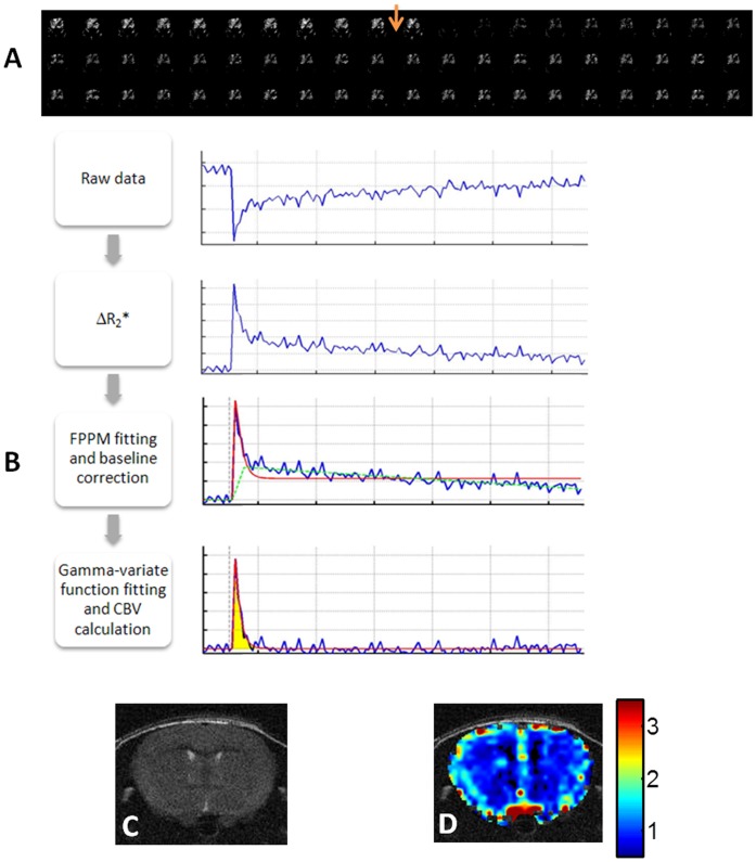
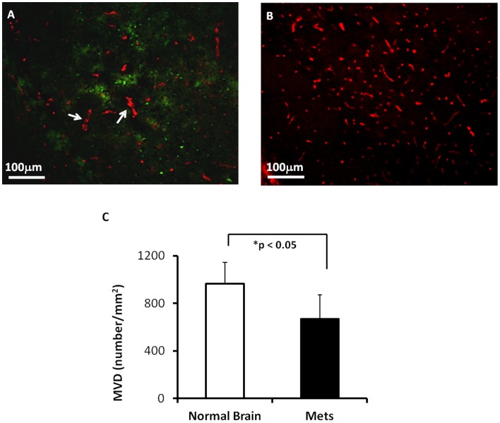
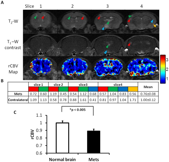
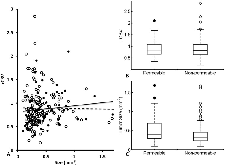
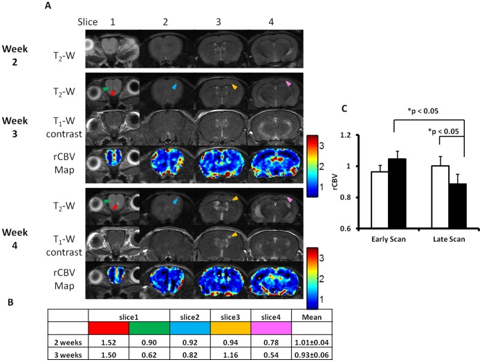
Longitudinal MRI was applied to monitor intracranial initiation and development of brain metastases and assess tumor vascular volume and permeability in a mouse model of breast cancer brain metastases. Using a 9.4T system, high resolution anatomic MRI and dynamic susceptibility contrast (DSC) perfusion MRI were acquired at different time points after an intracardiac injection of brain-tropic breast cancer MDA-MB231BR-EGFP cells. Three weeks post injection, multifocal brain metastases were first observed with hyperintensity on T2-weighted images, but isointensity on T1-weighted post contrast images, indicating that blood-tumor-barrier (BTB) at early stage of brain metastases was impermeable. Follow-up MRI revealed intracranial tumor growth and increased number of metastases that distributed throughout the whole brain. At the last scan on week 5, T1-weighted post contrast images detected BTB disruption in 160 (34%) of a total of 464 brain metastases. Enhancement in some of the metastases was only seen in partial regions of the tumor, suggesting intratumoral heterogeneity of BTB disruption. DSC MRI measurements of relative cerebral blood volume (rCBV) showed that rCBV of brain metastases was significantly lower (mean= 0.89±0.03) than that of contralateral normal brain (mean= 1.00±0.03; p<0.005). Intriguingly, longitudinal measurements revealed that rCBV of individual metastases at early stage was similar to, but became significantly lower than that of contralateral normal brain with tumor growth (p<0.05). The rCBV data were concordant with histological analysis of microvascular density (MVD). Moreover, comprehensive analysis suggested no significant correlation among tumor size, rCBV and BTB permeability. In conclusion, longitudinal MRI provides non-invasive in vivo assessments of spatial and temporal development of brain metastases and their vascular volume and permeability. The characteristic rCBV of brain metastases may have a diagnostic value.

摘要

采用纵向 MRI 监测颅内乳腺癌脑转移的起始和发展,并评估肿瘤血管容积和通透性。在心脏内注射嗜脑乳腺癌 MDA-MB231BR-EGFP 细胞后,在不同时间点使用 9.4T 系统获取高分辨率解剖 MRI 和动态对比敏感度(DSC)灌注 MRI。注射后 3 周,在 T2 加权图像上首次观察到多灶性脑转移,呈高信号,但 T1 加权对比后图像呈等信号,表明早期脑转移的血脑屏障(BTB)是不透的。后续 MRI 显示颅内肿瘤生长并增加了转移灶的数量,这些转移灶分布在整个大脑中。在第 5 周的最后一次扫描中,在总共 464 个脑转移灶中,有 160 个(34%)的 T1 加权对比后图像检测到 BTB 破坏。一些转移灶的增强仅在肿瘤的部分区域可见,这表明 BTB 破坏的肿瘤内异质性。DSC MRI 测量相对脑血容量(rCBV)显示,脑转移灶的 rCBV 明显低于对侧正常脑(rCBV=0.89±0.03)(rCBV=1.00±0.03;p<0.005)。有趣的是,纵向测量显示,早期单个转移灶的 rCBV 与肿瘤生长时对侧正常脑相似,但显著低于对侧正常脑(p<0.05)。rCBV 数据与微血管密度(MVD)的组织学分析一致。此外,综合分析表明肿瘤大小、rCBV 和 BTB 通透性之间没有显著相关性。总之,纵向 MRI 提供了脑转移及其血管容积和通透性的空间和时间发展的非侵入性体内评估。脑转移灶的特征性 rCBV 可能具有诊断价值。

https://cdn.ncbi.nlm.nih.gov/pmc/blobs/de74/3639286/2c6e8d13741b/pone.0062238.g006.jpg
https://cdn.ncbi.nlm.nih.gov/pmc/blobs/de74/3639286/f9c98f820aba/pone.0062238.g001.jpg
https://cdn.ncbi.nlm.nih.gov/pmc/blobs/de74/3639286/5ec1873e3c64/pone.0062238.g002.jpg
https://cdn.ncbi.nlm.nih.gov/pmc/blobs/de74/3639286/0d8c60ac17cf/pone.0062238.g003.jpg
https://cdn.ncbi.nlm.nih.gov/pmc/blobs/de74/3639286/101db7de7d50/pone.0062238.g004.jpg
https://cdn.ncbi.nlm.nih.gov/pmc/blobs/de74/3639286/07d603f0c75c/pone.0062238.g005.jpg
https://cdn.ncbi.nlm.nih.gov/pmc/blobs/de74/3639286/2c6e8d13741b/pone.0062238.g006.jpg

相似文献

1
Longitudinal MRI evaluation of intracranial development and vascular characteristics of breast cancer brain metastases in a mouse model.颅内乳腺癌脑转移瘤在小鼠模型中的纵向 MRI 评估:颅内发育和血管特征。
PLoS One. 2013 Apr 29;8(4):e62238. doi: 10.1371/journal.pone.0062238. Print 2013.
2
MRI evaluation of the effects of whole brain radiotherapy on breast cancer brain metastasis.MRI 评估全脑放疗对乳腺癌脑转移的影响。
Int J Radiat Biol. 2019 Mar;95(3):338-346. doi: 10.1080/09553002.2019.1554920. Epub 2019 Jan 7.
3
Potential for differentiation of pseudoprogression from true tumor progression with dynamic susceptibility-weighted contrast-enhanced magnetic resonance imaging using ferumoxytol vs. gadoteridol: a pilot study.使用 Ferumoxytol 与 Gadoteridol 行动态磁敏感对比增强磁共振成像对假性进展与真性肿瘤进展的鉴别潜能:一项初步研究。
Int J Radiat Oncol Biol Phys. 2011 Feb 1;79(2):514-23. doi: 10.1016/j.ijrobp.2009.10.072. Epub 2010 Apr 13.
4
Ultrasound imaging-guided intracardiac injection to develop a mouse model of breast cancer brain metastases followed by longitudinal MRI.超声成像引导下的心内注射以建立乳腺癌脑转移小鼠模型,随后进行纵向磁共振成像。
J Vis Exp. 2014 Mar 6(85):51146. doi: 10.3791/51146.
5
Dynamic susceptibility contrast (DSC) perfusion MRI in differential diagnosis between radionecrosis and neoangiogenesis in cerebral metastases using rCBV, rCBF and K2.动态对比磁共振灌注成像(DSC)在脑转移瘤放射性坏死与新生血管形成鉴别诊断中的应用:rCBV、rCBF 和 K2 的价值。
Radiol Med. 2018 Jul;123(7):545-552. doi: 10.1007/s11547-018-0866-7. Epub 2018 Mar 5.
6
Magnetic resonance imaging-based tumour perfusion parameters are biomarkers predicting response after radiation to brain metastases.基于磁共振成像的肿瘤灌注参数是预测脑转移瘤放疗后反应的生物标志物。
Clin Oncol (R Coll Radiol). 2014 Nov;26(11):704-12. doi: 10.1016/j.clon.2014.06.010. Epub 2014 Jul 10.
7
Correlation of relative permeability and relative cerebral blood volume in high-grade cerebral neoplasms.高级别脑肿瘤中相对渗透率与相对脑血容量的相关性
AJR Am J Roentgenol. 2006 Oct;187(4):1036-42. doi: 10.2214/AJR.04.0676.
8
Phase contrast MRI is an early marker of micrometastatic breast cancer development in the rat brain.磁共振相位对比成像可作为检测鼠脑乳腺癌微转移的早期标志物
NMR Biomed. 2012 May;25(5):726-36. doi: 10.1002/nbm.1786. Epub 2011 Sep 22.
9
Dynamic contrast-enhanced susceptibility-weighted perfusion imaging of intracranial tumors: a study using a 3T MR scanner.颅内肿瘤的动态对比增强磁共振血管造影灌注成像:一项使用3T磁共振扫描仪的研究
Diagn Interv Radiol. 2009 Mar;15(1):3-12.
10
Rat model of metastatic breast cancer monitored by MRI at 3 tesla and bioluminescence imaging with histological correlation.通过3特斯拉磁共振成像(MRI)和生物发光成像并结合组织学相关性监测的转移性乳腺癌大鼠模型
J Transl Med. 2009 Oct 20;7:88. doi: 10.1186/1479-5876-7-88.

引用本文的文献

1
Monitoring imatinib decreasing pericyte coverage and HIF-1α level in a colorectal cancer model by an ultrahigh-field multiparametric MRI approach.通过超高场多参数磁共振成像方法监测伊马替尼在结直肠癌模型中降低周细胞覆盖率和缺氧诱导因子-1α水平的情况。
J Transl Med. 2024 Jul 31;22(1):712. doi: 10.1186/s12967-024-05497-w.
2
Transfer Learning Approach to Vascular Permeability Changes in Brain Metastasis Post-Whole-Brain Radiotherapy.全脑放疗后脑转移瘤血管通透性变化的迁移学习方法
Cancers (Basel). 2023 May 10;15(10):2703. doi: 10.3390/cancers15102703.
3
Deep learning quantification of vascular pharmacokinetic parameters in mouse brain tumor models.

本文引用的文献

1
In vivo characterization of changing blood-tumor barrier permeability in a mouse model of breast cancer metastasis: a complementary magnetic resonance imaging approach.在乳腺癌转移的小鼠模型中,对血肿瘤屏障通透性变化的体内特征进行鉴定:一种互补的磁共振成像方法。
Invest Radiol. 2011 Nov;46(11):718-25. doi: 10.1097/RLI.0b013e318226c427.
2
The biology of brain metastases-translation to new therapies.脑转移瘤的生物学——向新疗法的转化。
Nat Rev Clin Oncol. 2011 Jun;8(6):344-56. doi: 10.1038/nrclinonc.2011.58. Epub 2011 Apr 12.
3
Brain metastases as preventive and therapeutic targets.
深度学习定量分析小鼠脑肿瘤模型中的血管药代动力学参数。
Front Biosci (Landmark Ed). 2022 Mar 16;27(3):99. doi: 10.31083/j.fbl2703099.
4
Preclinical Models of Brain Metastases in Breast Cancer.乳腺癌脑转移的临床前模型
Biomedicines. 2022 Mar 13;10(3):667. doi: 10.3390/biomedicines10030667.
5
Current landscape and future perspectives in preclinical MR and PET imaging of brain metastasis.脑转移瘤临床前磁共振成像和正电子发射断层显像的现状与未来展望
Neurooncol Adv. 2021 Oct 14;3(1):vdab151. doi: 10.1093/noajnl/vdab151. eCollection 2021 Jan-Dec.
6
Novel human melanoma brain metastasis models in athymic nude fox1 mice: Site-specific metastasis patterns reflecting their clinical origin.新型无胸腺裸鼠人黑色素瘤脑转移模型:具有临床起源特征的特定部位转移模式。
Cancer Med. 2021 Dec;10(23):8604-8613. doi: 10.1002/cam4.4334. Epub 2021 Oct 6.
7
MRI evaluation of the effects of whole brain radiotherapy on breast cancer brain metastasis.MRI 评估全脑放疗对乳腺癌脑转移的影响。
Int J Radiat Biol. 2019 Mar;95(3):338-346. doi: 10.1080/09553002.2019.1554920. Epub 2019 Jan 7.
8
EPMA position paper in cancer: current overview and future perspectives.电子探针微量分析在癌症领域的立场文件:当前概述与未来展望
EPMA J. 2015 Apr 15;6(1):9. doi: 10.1186/s13167-015-0030-6. eCollection 2015.
9
Ultrasound imaging-guided intracardiac injection to develop a mouse model of breast cancer brain metastases followed by longitudinal MRI.超声成像引导下的心内注射以建立乳腺癌脑转移小鼠模型,随后进行纵向磁共振成像。
J Vis Exp. 2014 Mar 6(85):51146. doi: 10.3791/51146.
10
Ex vivo Evans blue assessment of the blood brain barrier in three breast cancer brain metastasis models.三种乳腺癌脑转移模型中血脑屏障的 Evans 蓝体外评估。
Breast Cancer Res Treat. 2014 Feb;144(1):93-101. doi: 10.1007/s10549-014-2854-5. Epub 2014 Feb 8.
脑转移作为预防和治疗的靶点。
Nat Rev Cancer. 2011 May;11(5):352-63. doi: 10.1038/nrc3053. Epub 2011 Apr 7.
4
Mouse models of advanced spontaneous metastasis for experimental therapeutics.用于实验治疗的晚期自发性转移的小鼠模型。
Nat Rev Cancer. 2011 Feb;11(2):135-41. doi: 10.1038/nrc3001.
5
Heterogeneous blood-tumor barrier permeability determines drug efficacy in experimental brain metastases of breast cancer.异质性血脑屏障通透性决定乳腺癌实验性脑转移的药物疗效。
Clin Cancer Res. 2010 Dec 1;16(23):5664-78. doi: 10.1158/1078-0432.CCR-10-1564. Epub 2010 Sep 9.
6
Cancer statistics, 2010.癌症统计数据,2010 年。
CA Cancer J Clin. 2010 Sep-Oct;60(5):277-300. doi: 10.3322/caac.20073. Epub 2010 Jul 7.
7
Capturing changes in the brain microenvironment during initial steps of breast cancer brain metastasis.捕获乳腺癌脑转移初始阶段大脑微环境的变化。
Am J Pathol. 2010 Jun;176(6):2958-71. doi: 10.2353/ajpath.2010.090838. Epub 2010 Apr 9.
8
Real-time imaging reveals the single steps of brain metastasis formation.实时成像揭示了脑转移形成的单个步骤。
Nat Med. 2010 Jan;16(1):116-22. doi: 10.1038/nm.2072. Epub 2009 Dec 20.
9
Rat model of metastatic breast cancer monitored by MRI at 3 tesla and bioluminescence imaging with histological correlation.通过3特斯拉磁共振成像(MRI)和生物发光成像并结合组织学相关性监测的转移性乳腺癌大鼠模型
J Transl Med. 2009 Oct 20;7:88. doi: 10.1186/1479-5876-7-88.
10
The vascular basement membrane as "soil" in brain metastasis.血管基底膜作为脑转移中的“土壤”
PLoS One. 2009 Jun 10;4(6):e5857. doi: 10.1371/journal.pone.0005857.