INSERM U1016, Institut Cochin, Université Paris Descartes, 24 rue du Faubourg Saint-Jacques, F75014 Paris, France.
PLoS One. 2013 Apr 25;8(4):e62919. doi: 10.1371/journal.pone.0062919. Print 2013.
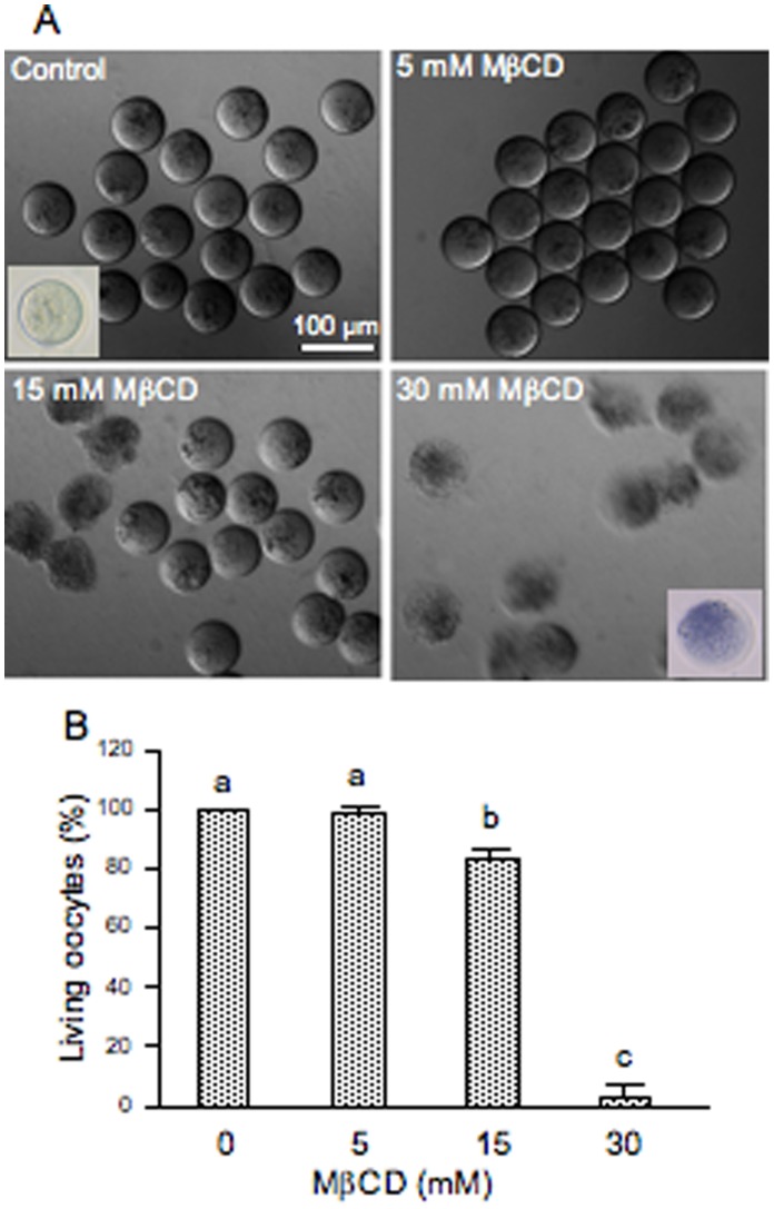
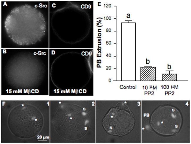
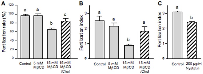
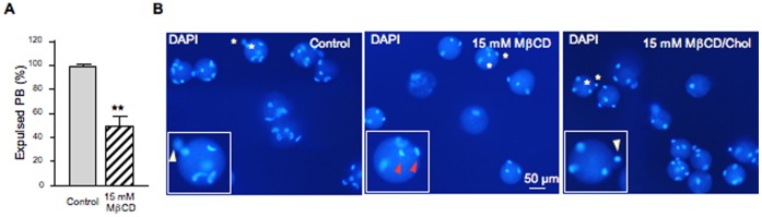
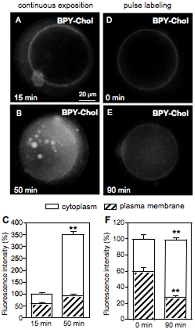
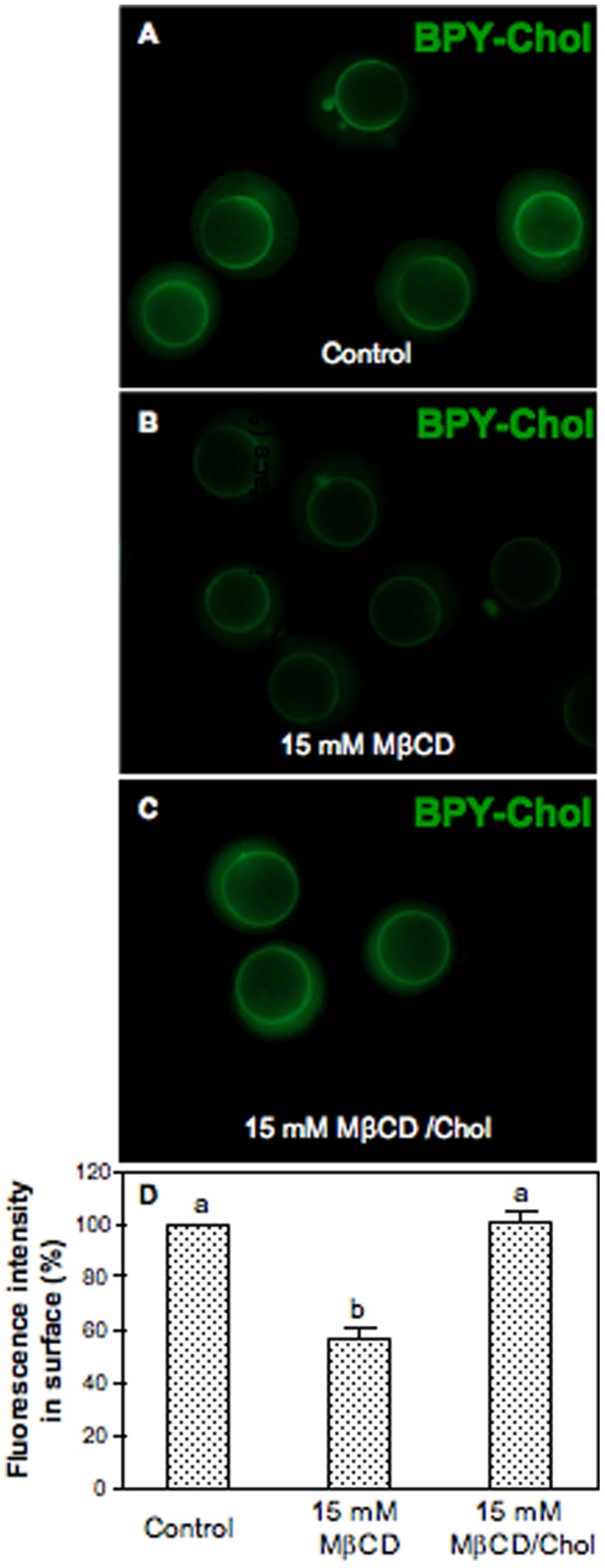
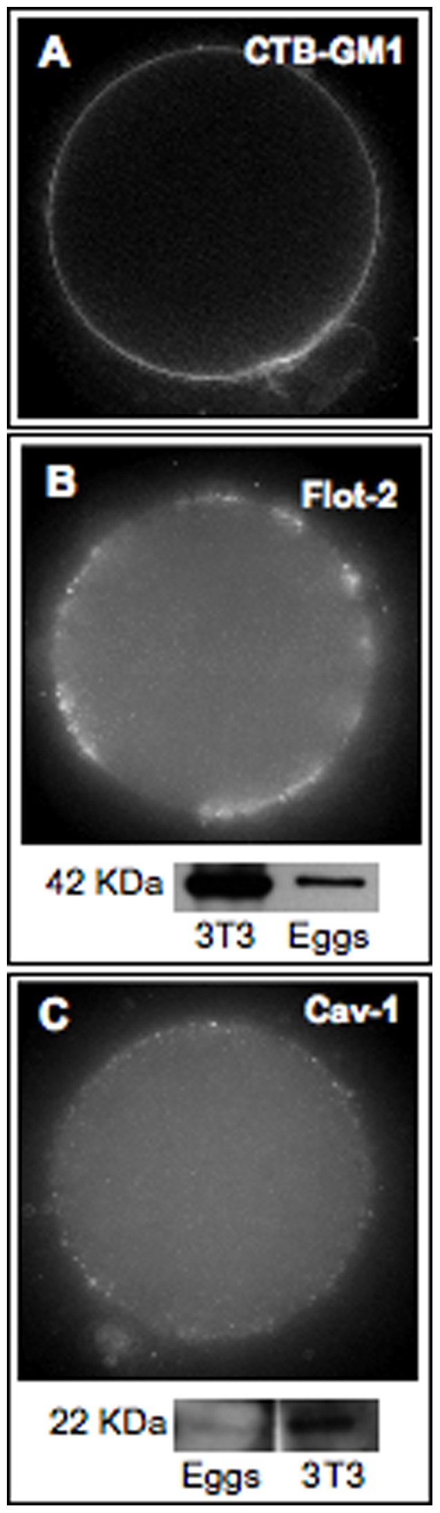
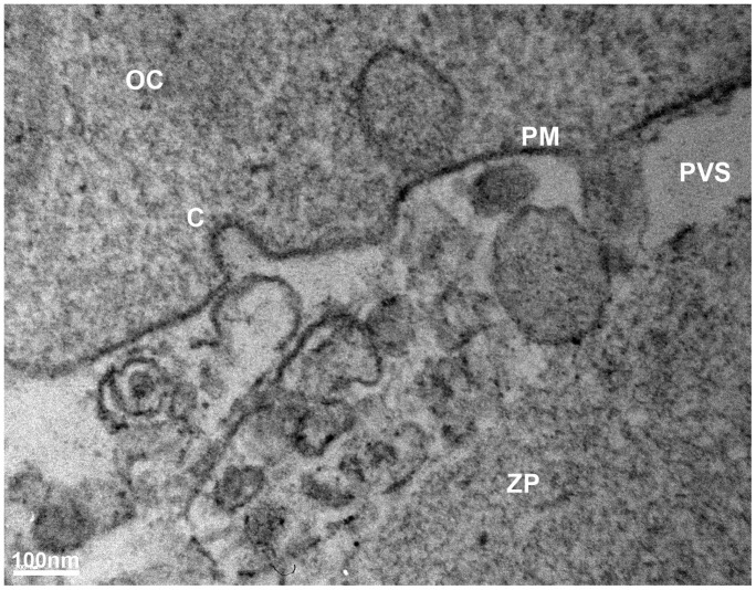
Drastic membrane reorganization occurs when mammalian sperm binds to and fuses with the oocyte membrane. Two oocyte protein families are essential for fertilization, tetraspanins and glycosylphosphatidylinositol-anchored proteins. The firsts are associated to tetraspanin-enriched microdomains and the seconds to lipid rafts. Here we report membrane raft involvement in mouse fertilization assessed by cholesterol modulation using methyl-β-cyclodextrin. Cholesterol removal induced: (1) a decrease of the fertilization rate and index; and (2) a delay in the extrusion of the second polar body. Cholesterol repletion recovered the fertilization ability of cholesterol-depleted oocytes, indicating reversibility of these effects. In vivo time-lapse analyses using fluorescent cholesterol permitted to identify the time-point at which the probe is mainly located at the plasma membrane enabling the estimation of the extent of the cholesterol depletion. We confirmed that the mouse oocyte is rich in rafts according to the presence of the raft marker lipid, ganglioside GM1 on the membrane of living oocytes and we identified the coexistence of two types of microdomains, planar rafts and caveolae-like structures, by terms of two differential rafts markers, flotillin-2 and caveolin-1, respectively. Moreover, this is the first report that shows characteristic caveolae-like invaginations in the mouse oocyte identified by electron microscopy. Raft disruption by cholesterol depletion disturbed the subcellular localization of the signal molecule c-Src and the inhibition of Src kinase proteins prevented second polar body extrusion, consistent with a role of Src-related kinases in fertilization via signaling complexes. Our data highlight the functional importance of intact membrane rafts for mouse fertilization and its dependence on cholesterol.
哺乳动物精子与卵母细胞膜结合并融合时,会发生剧烈的膜重排。两种卵母细胞蛋白家族对于受精是必不可少的,即四跨膜蛋白和糖基磷脂酰肌醇锚定蛋白。前者与富含四跨膜蛋白的微区相关,后者与脂筏相关。在这里,我们通过使用甲基-β-环糊精调节胆固醇来评估膜筏在小鼠受精中的作用。胆固醇去除:(1)降低受精率和指数;(2)延迟第二极体的排出。胆固醇补充恢复了胆固醇耗尽卵母细胞的受精能力,表明这些效应是可逆的。使用荧光胆固醇进行的体内延时分析允许确定探针主要位于质膜的时间点,从而可以估计胆固醇耗尽的程度。我们通过存在于活卵母细胞膜上的筏标记脂质神经节苷脂 GM1 证实了小鼠卵母细胞富含筏,并且通过两种不同的筏标记物,分别是 flotillin-2 和 caveolin-1,鉴定了两种类型的微区,即平面筏和类似小窝的结构的共存。此外,这是首次报道显示在电子显微镜下鉴定的小鼠卵母细胞中存在特征性的类似小窝内陷。胆固醇耗尽破坏了信号分子 c-Src 的亚细胞定位,Src 激酶蛋白的抑制阻止了第二极体的排出,这与 Src 相关激酶通过信号复合物在受精中的作用一致。我们的数据强调了完整的膜筏对于小鼠受精的功能重要性及其对胆固醇的依赖性。