Ramaswamy Anikode Subramanian, Manjunatha Hanumanthappa Krishnappa, Sunilkumar Bylappa, Arunkumar Sulkunte Palaksha
Department of Pathology, P.E.S Institute of Medical Sciences and Research, Kuppam, Andhra Pradesh, India.
N Am J Med Sci. 2013 Feb;5(2):124-8. doi: 10.4103/1947-2714.107532.
Cysts of the skin are one of the commonly excised specimens in the surgical outpatient department. A majority of them being clinically diagnosed as sebaceous cysts, their true nature is only discernible on histopathological examination. Closer examination of the type of keratinization involved will throw light into the exact nature of the cyst. Trichilemmal or Pilar cyst is one such entity, which presents in both a non-neoplastic and neoplastic form.
The present retrospective observational study was undertaken to find out the incidence of these cysts in surgical pathology practice in a rural hospital and to enlist the various morphological forms that these cysts may take.
The histopathology files were reviewed for a period of 6 years for cases coded as pilar cyst.
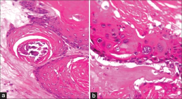
A total of eight cases (5.75%) were identified, which showed features of trichilemmal differentiation. A single case each of proliferating trichilemmal cyst and malignant proliferating trichilemmal tumors were noted. Most of the cases were seen among females on the scalp.
Trichilemmal tumor is an uncommon histopathological entity. Many of these lesions may be mistakenly diagnosed due to lack of recognition of the unique type of keratinization.
皮肤囊肿是外科门诊常见的切除标本之一。它们大多在临床上被诊断为皮脂腺囊肿,其真实性质只有在组织病理学检查中才能辨别。仔细检查所涉及的角化类型将有助于明确囊肿的确切性质。毛囊囊肿就是这样一种实体,它以非肿瘤性和肿瘤性两种形式出现。
本回顾性观察研究旨在查明农村医院外科病理学实践中这些囊肿的发病率,并列出这些囊肿可能呈现的各种形态学形式。
回顾组织病理学档案6年中编码为毛囊囊肿的病例。
共识别出8例(5.75%),表现出毛囊分化特征。记录到1例增殖性毛囊囊肿和1例恶性增殖性毛囊肿瘤。大多数病例见于头皮的女性。
毛囊肿瘤是一种罕见的组织病理学实体。由于对独特的角化类型认识不足,许多这些病变可能被误诊。