Department of Physiology, Michigan State University, East Lansing, MI 48824, USA.
Toxicol Appl Pharmacol. 2013 Aug 15;271(1):20-9. doi: 10.1016/j.taap.2013.04.018. Epub 2013 Apr 30.
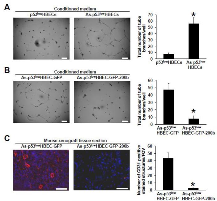
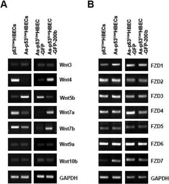
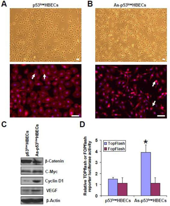
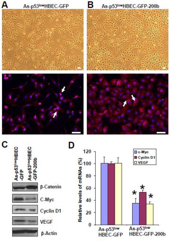
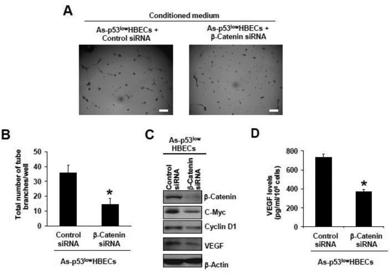
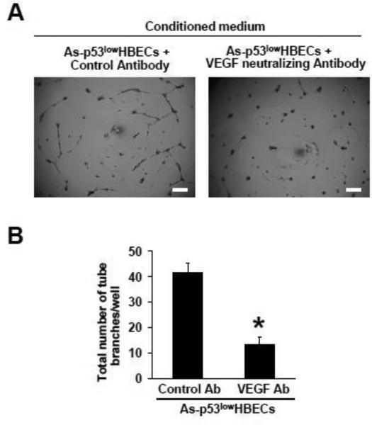
Arsenic exposure represents a major health concern increasing cancer risks, yet the mechanism of arsenic carcinogenesis has not been elucidated. We and others recently reported that cell malignant transformation by arsenic is accompanied by epithelial to mesenchymal transition (EMT). However, the role of EMT in arsenic carcinogenesis is not well understood. Although previous studies showed that short term exposure of endothelial cells to arsenic stimulated angiogenesis, it remains to be determined whether cells that were malignantly transformed by long term arsenic exposure have a pro-angiogenic effect. The objective of this study was to investigate the effect of arsenic-transformed human bronchial epithelial cells that underwent EMT on angiogenesis and the underlying mechanism. It was found that the conditioned medium from arsenic-transformed cells strongly stimulated tube formation by human umbilical vein endothelial cells (HUVECs). Moreover, enhanced angiogenesis was detected in mouse xenograft tumor tissues resulting from inoculation of arsenic-transformed cells. Mechanistic studies revealed that β-catenin was activated in arsenic-transformed cells up-regulating its target gene expression including angiogenic-stimulating vascular endothelial growth factor (VEGF). Stably expressing microRNA-200b in arsenic-transformed cells that reversed EMT inhibited β-catenin activation, decreased VEGF expression and reduced tube formation by HUVECs. SiRNA knockdown β-catenin decreased VEGF expression. Adding a VEGF neutralizing antibody into the conditioned medium from arsenic-transformed cells impaired tube formation by HUVECs. Reverse transcriptase-PCR analysis revealed that the mRNA levels of canonical Wnt ligands were not increased in arsenic-transformed cells. These findings suggest that EMT in arsenic-transformed cells promotes angiogenesis through activating β-catenin-VEGF pathway.
砷暴露是一个主要的健康问题,会增加癌症风险,但砷致癌的机制尚未阐明。我们和其他人最近报道,砷引起的细胞恶性转化伴随着上皮间质转化(EMT)。然而,EMT 在砷致癌中的作用尚不清楚。虽然先前的研究表明,内皮细胞短期暴露于砷会刺激血管生成,但仍需要确定长期暴露于砷的恶性转化细胞是否具有促血管生成作用。本研究的目的是研究 EMT 的砷转化人支气管上皮细胞对血管生成的影响及其潜在机制。结果发现,砷转化细胞的条件培养基强烈刺激人脐静脉内皮细胞(HUVEC)的管形成。此外,接种砷转化细胞的小鼠异种移植肿瘤组织中检测到增强的血管生成。机制研究表明,β-连环蛋白在砷转化细胞中被激活,上调其靶基因表达,包括血管生成刺激血管内皮生长因子(VEGF)。稳定表达 microRNA-200b 可逆转 EMT,抑制β-连环蛋白激活,降低 VEGF 表达,减少 HUVEC 的管形成。siRNA 敲低β-连环蛋白可降低 VEGF 表达。向砷转化细胞的条件培养基中加入 VEGF 中和抗体可损害 HUVEC 的管形成。逆转录-PCR 分析显示,砷转化细胞中经典 Wnt 配体的 mRNA 水平没有增加。这些发现表明,砷转化细胞中的 EMT 通过激活β-连环蛋白-VEGF 通路促进血管生成。