Neogi Tuhina, Bowes Michael A, Niu Jingbo, De Souza Kevin M, Vincent Graham R, Goggins Joyce, Zhang Yuqing, Felson David T
Boston University School of Medicine, Boston, MA 02118, USA.
Arthritis Rheum. 2013 Aug;65(8):2048-58. doi: 10.1002/art.37987.
To examine whether magnetic resonance imaging (MRI)-based 3-dimensional (3-D) bone shape predicts the onset of radiographic knee osteoarthritis (OA).
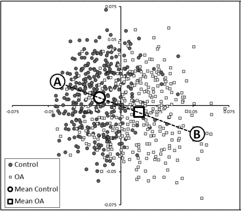
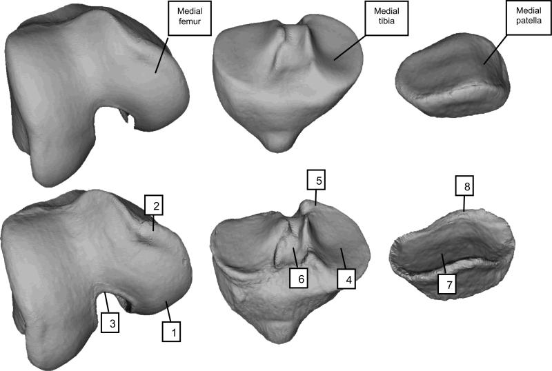
We conducted a case-control study using data from the Osteoarthritis Initiative by identifying knees that developed incident tibiofemoral radiographic knee OA (case knees) during followup, and matching them each to 2 random control knees. Using knee MRIs, we performed active appearance modeling of the femur, tibia, and patella and linear discriminant analysis to identify vectors that best classified knees with OA versus those without OA. Vectors were scaled such that -1 and +1 represented the mean non-OA and mean OA shapes, respectively. We examined the relation of 3-D bone shape to incident OA (new-onset Kellgren and Lawrence [K/L] grade ≥2) occurring 12 months later using conditional logistic regression.
A total of 178 case knees (incident OA) were matched to 353 control knees. The whole joint (i.e., tibia, femur, and patella) 3-D bone shape vector had the strongest magnitude of effect, with knees in the highest tertile having a 3.0 times higher likelihood of developing incident radiographic knee OA 12 months later compared with those in the lowest tertile (95% confidence interval [95% CI] 1.8-5.0, P < 0.0001). The associations were even stronger among knees that had completely normal radiographs before incidence (K/L grade of 0) (odds ratio 12.5 [95% CI 4.0-39.3]). Bone shape at baseline, often several years before incidence, predicted later OA.
MRI-based 3-D bone shape predicted the later onset of radiographic OA. Further study is warranted to determine whether such methods can detect treatment effects in trials and provide insight into the pathophysiology of OA development.
探讨基于磁共振成像(MRI)的三维(3-D)骨形态是否能预测膝关节X线骨关节炎(OA)的发病。
我们利用骨关节炎倡议组织的数据进行了一项病例对照研究,通过识别随访期间发生胫股关节X线膝关节OA(病例膝关节)的膝关节,并将每个病例膝关节与2个随机对照膝关节进行匹配。利用膝关节MRI,我们对股骨、胫骨和髌骨进行了主动外观建模,并进行线性判别分析,以识别能将OA膝关节与非OA膝关节最佳分类的向量。对向量进行缩放,使-1和+1分别代表平均非OA和平均OA形态。我们使用条件逻辑回归分析了3-D骨形态与12个月后发生的新发OA(新出现的Kellgren和Lawrence [K/L]分级≥2)之间的关系。
共178个病例膝关节(新发OA)与353个对照膝关节匹配。整个关节(即胫骨、股骨和髌骨)的3-D骨形态向量具有最强的效应量,处于最高三分位数的膝关节在12个月后发生新发X线膝关节OA的可能性是处于最低三分位数膝关节的3.0倍(95%置信区间[95% CI] 1.8-5.0,P < 0.0001)。在发病前X线片完全正常(K/L分级为0)的膝关节中,这种关联更强(优势比12.5 [95% CI 4.0-39.3])。发病前数年的基线骨形态可预测后期的OA。
基于MRI的3-D骨形态可预测后期X线OA的发病。有必要进一步研究以确定此类方法是否能在试验中检测到治疗效果,并深入了解OA发展的病理生理学。