Institute of Inorganic and Analytical Chemistry, Justus Liebig University, Schubertstrasse 60, 35392 Giessen, Germany.
Histochem Cell Biol. 2013 Jun;139(6):759-83. doi: 10.1007/s00418-013-1097-6. Epub 2013 May 8.
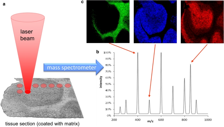
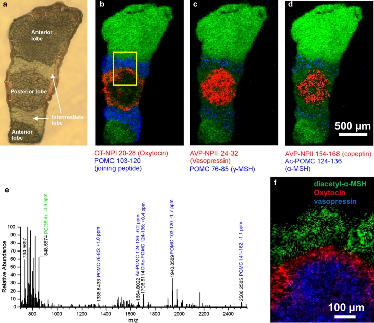
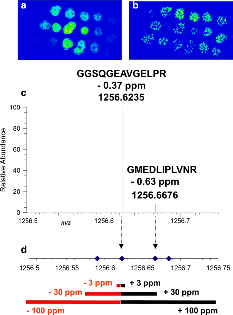
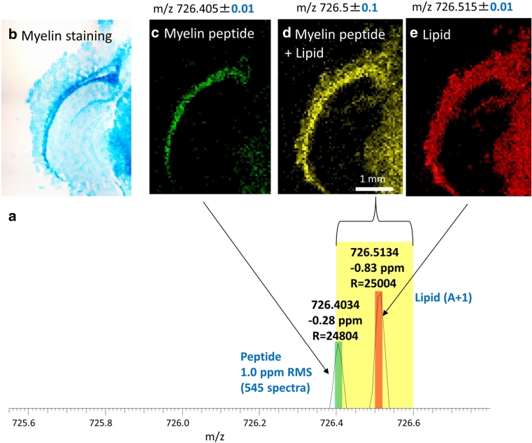
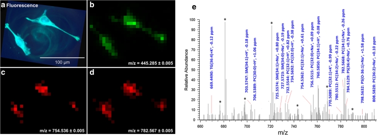
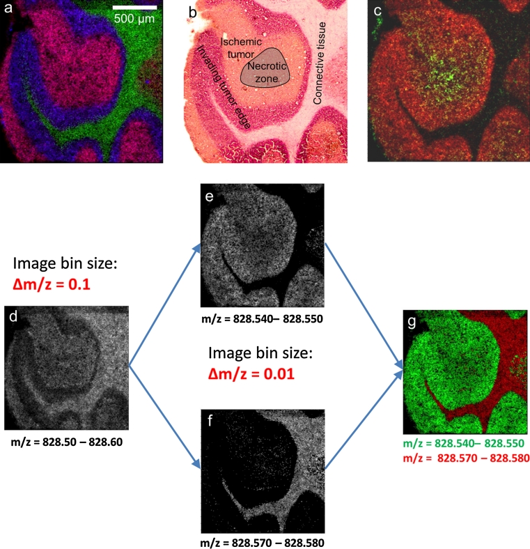
Mass spectrometry (MS) imaging links molecular information and the spatial distribution of analytes within a sample. In contrast to most histochemical techniques, mass spectrometry imaging can differentiate molecular modifications and does not require labeling of targeted compounds. We have recently introduced the first mass spectrometry imaging method that provides highly specific molecular information (high resolution and accuracy in mass) at cellular dimensions (high resolution in space). This method is based on a matrix-assisted laser desorption/ionization (MALDI) imaging source working at atmospheric pressure which is coupled to an orbital trapping mass spectrometer. Here, we present a number of application examples and demonstrate the benefit of 'mass spectrometry imaging with high resolution in mass and space.' Phospholipids, peptides and drug compounds were imaged in a number of tissue samples at a spatial resolution of 5-10 μm. Proteins were analyzed after on-tissue tryptic digestion at 50-μm resolution. Additional applications include the analysis of single cells and of human lung carcinoma tissue as well as the first MALDI imaging measurement of tissue at 3 μm pixel size. MS image analysis for all these experiments showed excellent correlation with histological staining evaluation. The high mass resolution (R = 30,000) and mass accuracy (typically 1 ppm) proved to be essential for specific image generation and reliable identification of analytes in tissue samples. The ability to combine the required high-quality mass analysis with spatial resolution in the range of single cells is a unique feature of our method. With that, it has the potential to supplement classical histochemical protocols and to provide new insights about molecular processes on the cellular level.
质谱成像(MSI)将分子信息与样品中分析物的空间分布联系起来。与大多数组织化学技术不同,质谱成像可以区分分子修饰,并且不需要对靶向化合物进行标记。我们最近引入了第一种提供高分辨率(高空间分辨率)和高特异性分子信息(高质量分辨率和准确性)的质谱成像方法。该方法基于在大气压下工作的基质辅助激光解吸/电离(MALDI)成像源,与轨道俘获质谱仪耦合。在这里,我们展示了一些应用实例,并证明了“具有高质量分辨率和空间分辨率的质谱成像”的优势。在一些组织样本中,以 5-10μm 的空间分辨率成像了磷脂、肽和药物化合物。在 50-μm 的分辨率下进行组织内胰蛋白酶消化后分析蛋白质。其他应用包括单细胞分析和人肺癌组织分析,以及 3μm 像素大小的组织的第一个 MALDI 成像测量。所有这些实验的 MS 图像分析均显示与组织学染色评估具有出色的相关性。高质量分辨率(R=30,000)和质量准确性(通常为 1ppm)对于特定图像生成和可靠识别组织样本中的分析物至关重要。将所需的高质量分析与单细胞范围内的空间分辨率相结合的能力是我们方法的独特特征。它有可能补充经典的组织化学方案,并提供关于细胞水平上分子过程的新见解。