• 文献检索
  • 文档翻译
  • 深度研究
  • 学术资讯
  • Suppr Zotero 插件Zotero 插件
  • 邀请有礼
  • 套餐&价格
  • 历史记录
应用&插件
Suppr Zotero 插件Zotero 插件浏览器插件Mac 客户端Windows 客户端微信小程序
定价
高级版会员购买积分包购买API积分包
服务
文献检索文档翻译深度研究API 文档MCP 服务
关于我们
关于 Suppr公司介绍联系我们用户协议隐私条款
关注我们

Suppr 超能文献

核心技术专利:CN118964589B侵权必究
粤ICP备2023148730 号-1Suppr @ 2026

文献检索

告别复杂PubMed语法,用中文像聊天一样搜索,搜遍4000万医学文献。AI智能推荐,让科研检索更轻松。

立即免费搜索

文件翻译

保留排版,准确专业,支持PDF/Word/PPT等文件格式,支持 12+语言互译。

免费翻译文档

深度研究

AI帮你快速写综述,25分钟生成高质量综述,智能提取关键信息,辅助科研写作。

立即免费体验

评估基于计算机辅助纹理分析的工具在 DCE-MR 乳腺摄影可疑病变的参数图像中的应用可行性。

Assessment of feasibility to use computer aided texture analysis based tool for parametric images of suspicious lesions in DCE-MR mammography.

机构信息

Bilecik Şeyh Edebali University, Turkey.

出版信息

Comput Math Methods Med. 2013;2013:872676. doi: 10.1155/2013/872676. Epub 2013 Apr 9.

DOI:10.1155/2013/872676
PMID:23653668
原文链接:https://pmc.ncbi.nlm.nih.gov/articles/PMC3638704/
Abstract

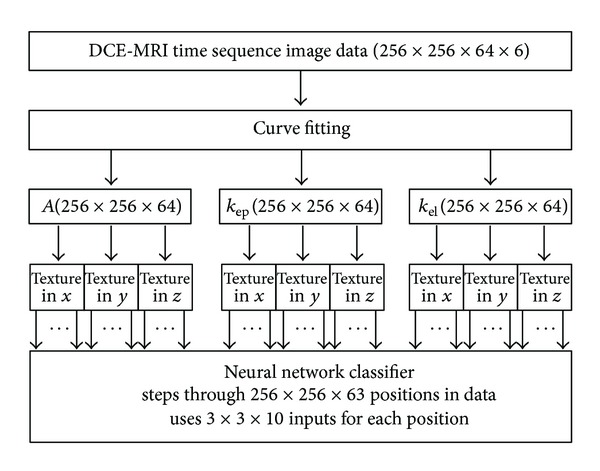
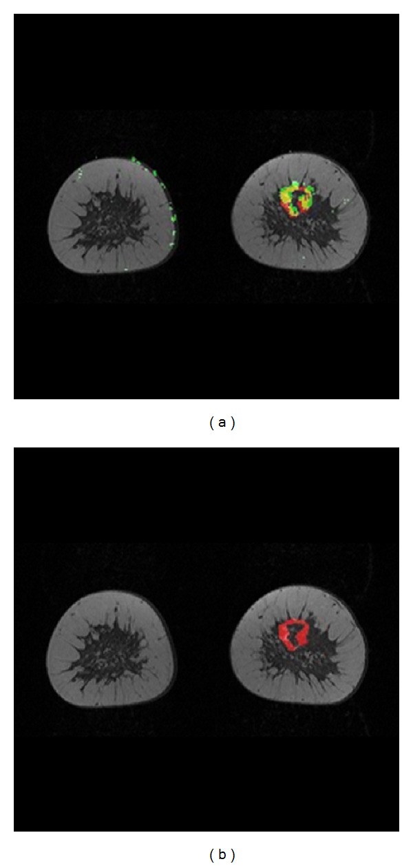
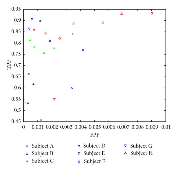
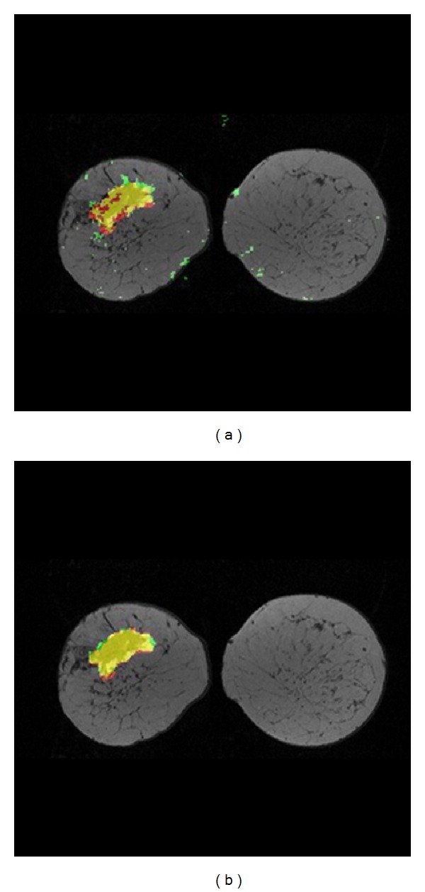
Our aim was to analyze the feasibility of computer aided malignant tumor detection using the traditional texture analysis applied on two-compartment-based parameter pseudoimages of dynamic contrast-enhanced magnetic resonance (DCE-MR) breast image data. A major contribution of this research will be the through-plane assessment capability. Texture analysis was performed on two-compartment-based pseudo images of DCE-MRI datasets of breast data of eight subjects. The resulting texture parameter pseudo images were inputted to a feedforward neural network classification system which uses the manual segmentations of a primary radiologist as a gold standard, and each voxel was assigned as malignant or nonmalignant. The classification results were compared with the lesions manually segmented by a second radiologist. Results show that the mean true positive fraction (TPF) and false positive fraction (FPF) performance of the classifier vs. primary radiologist is statistically as good as the mean TPF and FPF performance of the second radiologist vs. primary radiologist with a confidence interval of 95% using a one-sample t-test with α = 0.05. In the experiment implemented on all of the eight subjects, all malignant tumors marked by the primary radiologist were classified to be malignant by the computer classifier. Our results have shown that neural network classification using the textural parameters for automated screening of two-compartment-based parameter pseudo images of DCE-MRI as input data can be a supportive tool for the radiologists in the preassessment stage to show the possible cancerous regions and in the postassessment stage to review the segmentations especially in analyzing complex DCE-MRI cases.

摘要

我们的目的是分析使用传统的纹理分析在基于双室的动态对比增强磁共振(DCE-MR)乳腺图像数据的参数伪影上检测恶性肿瘤的可行性。这项研究的一个主要贡献将是贯穿平面评估能力。对 8 例受试者的 DCE-MRI 数据集的双室基于伪影的乳腺数据进行了纹理分析。将得到的纹理参数伪影输入到前馈神经网络分类系统中,该系统使用初级放射科医生的手动分割作为金标准,将每个体素分配为恶性或非恶性。将分类结果与第二位放射科医生手动分割的病变进行比较。结果表明,与初级放射科医生相比,分类器的平均真阳性分数(TPF)和假阳性分数(FPF)性能与第二位放射科医生与初级放射科医生相比的平均 TPF 和 FPF 性能具有统计学意义,置信区间为 95%使用单侧 t 检验,α = 0.05。在对所有 8 名受试者进行的实验中,计算机分类器将初级放射科医生标记的所有恶性肿瘤均分类为恶性。我们的结果表明,使用纹理参数对基于双室的 DCE-MRI 参数伪影进行神经网络分类作为输入数据,可以成为放射科医生在预评估阶段显示可能的癌症区域的辅助工具,以及在评估阶段审查分割的工具,特别是在分析复杂的 DCE-MRI 病例时。

https://cdn.ncbi.nlm.nih.gov/pmc/blobs/f746/3638704/9ca7991672ee/CMMM2013-872676.004.jpg
https://cdn.ncbi.nlm.nih.gov/pmc/blobs/f746/3638704/c1f8659e23c4/CMMM2013-872676.001.jpg
https://cdn.ncbi.nlm.nih.gov/pmc/blobs/f746/3638704/22ef663cc1c1/CMMM2013-872676.002.jpg
https://cdn.ncbi.nlm.nih.gov/pmc/blobs/f746/3638704/12db1e8bebab/CMMM2013-872676.003.jpg
https://cdn.ncbi.nlm.nih.gov/pmc/blobs/f746/3638704/9ca7991672ee/CMMM2013-872676.004.jpg

相似文献

1
Assessment of feasibility to use computer aided texture analysis based tool for parametric images of suspicious lesions in DCE-MR mammography.评估基于计算机辅助纹理分析的工具在 DCE-MR 乳腺摄影可疑病变的参数图像中的应用可行性。
Comput Math Methods Med. 2013;2013:872676. doi: 10.1155/2013/872676. Epub 2013 Apr 9.
2
Malignant-lesion segmentation using 4D co-occurrence texture analysis applied to dynamic contrast-enhanced magnetic resonance breast image data.将4D共生纹理分析应用于动态对比增强磁共振乳腺图像数据的恶性病变分割
J Magn Reson Imaging. 2007 Mar;25(3):495-501. doi: 10.1002/jmri.20837.
3
Multimodality computer-aided breast cancer diagnosis with FFDM and DCE-MRI.基于 FFDM 和 DCE-MRI 的多模态计算机辅助乳腺癌诊断。
Acad Radiol. 2010 Sep;17(9):1158-67. doi: 10.1016/j.acra.2010.04.015.
4
Textural kinetics: a novel dynamic contrast-enhanced (DCE)-MRI feature for breast lesion classification.纹理动力学:一种新的动态对比增强(DCE)MRI 特征,用于乳腺病变分类。
J Digit Imaging. 2011 Jun;24(3):446-63. doi: 10.1007/s10278-010-9298-1.
5
Automatic identification and classification of characteristic kinetic curves of breast lesions on DCE-MRI.动态对比增强磁共振成像(DCE-MRI)中乳腺病变特征性动力学曲线的自动识别与分类
Med Phys. 2006 Aug;33(8):2878-87. doi: 10.1118/1.2210568.
6
Multispectral co-occurrence with three random variables in dynamic contrast enhanced magnetic resonance imaging of breast cancer.乳腺癌动态对比增强磁共振成像中三个随机变量的多光谱共生。
IEEE Trans Med Imaging. 2008 Oct;27(10):1425-31. doi: 10.1109/TMI.2008.922181.
7
Task-based assessment of a convolutional neural network for segmenting breast lesions for radiomic analysis.基于任务的卷积神经网络在乳腺病变分割中的放射组学分析评估。
Magn Reson Med. 2019 Aug;82(2):786-795. doi: 10.1002/mrm.27758. Epub 2019 Apr 8.
8
Potential of computer-aided diagnosis of high spectral and spatial resolution (HiSS) MRI in the classification of breast lesions.高光谱和空间分辨率(HiSS)MRI 计算机辅助诊断在乳腺病变分类中的应用潜力。
J Magn Reson Imaging. 2014 Jan;39(1):59-67. doi: 10.1002/jmri.24145. Epub 2013 Sep 10.
9
Multilevel analysis of spatiotemporal association features for differentiation of tumor enhancement patterns in breast DCE-MRI.基于多时相分析的乳腺 DCE-MRI 肿瘤增强模式鉴别特征研究。
Med Phys. 2010 Aug;37(8):3940-56. doi: 10.1118/1.3446799.
10
Characterization of spatiotemporal changes for the classification of dynamic contrast-enhanced magnetic-resonance breast lesions.动态对比增强磁共振乳腺病变分类的时空变化特征。
Artif Intell Med. 2013 Jun;58(2):101-14. doi: 10.1016/j.artmed.2013.03.002. Epub 2013 Mar 30.

引用本文的文献

1
Using Deep Neural Network Approach for Multiple-Class Assessment of Digital Mammography.使用深度神经网络方法进行数字乳腺摄影的多类别评估。
Healthcare (Basel). 2022 Nov 27;10(12):2382. doi: 10.3390/healthcare10122382.
2
A novel framework for evaluating the image accuracy of dynamic MRI and the application on accelerated breast DCE MRI.一种用于评估动态磁共振成像(MRI)图像准确性的新型框架及其在乳腺动态对比增强MRI加速成像中的应用。
MAGMA. 2018 Apr;31(2):309-320. doi: 10.1007/s10334-017-0648-6. Epub 2017 Sep 11.
3
Application of Texture Analysis to Study Small Vessel Disease and Blood-Brain Barrier Integrity.

本文引用的文献

1
A theoretical comparison of texture algorithms.纹理算法的理论比较。
IEEE Trans Pattern Anal Mach Intell. 1980 Mar;2(3):204-22. doi: 10.1109/tpami.1980.4767008.
2
Texture anisotropy in 3-D images.三维图像中的纹理各向异性。
IEEE Trans Image Process. 1999;8(3):346-60. doi: 10.1109/83.748890.
3
An adaptive tissue characterization network for model-free visualization of dynamic contrast-enhanced magnetic resonance image data.一种用于动态对比增强磁共振图像数据无模型可视化的自适应组织表征网络。
纹理分析在研究小血管疾病和血脑屏障完整性中的应用。
Front Neurol. 2017 Jul 19;8:327. doi: 10.3389/fneur.2017.00327. eCollection 2017.
4
Wavelet Entropy and Directed Acyclic Graph Support Vector Machine for Detection of Patients with Unilateral Hearing Loss in MRI Scanning.小波熵与有向无环图支持向量机用于在MRI扫描中检测单侧听力损失患者
Front Comput Neurosci. 2016 Oct 19;10:106. doi: 10.3389/fncom.2016.00106. eCollection 2016.
IEEE Trans Med Imaging. 2005 Oct;24(10):1256-66. doi: 10.1109/TMI.2005.854517.
4
Classification of signal-time curves obtained by dynamic magnetic resonance mammography: statistical comparison of quantitative methods.动态磁共振乳腺成像获得的信号-时间曲线分类:定量方法的统计比较
Invest Radiol. 2005 Jul;40(7):442-7. doi: 10.1097/01.rli.0000164788.73298.ae.
5
Neural network approach to the segmentation and classification of dynamic magnetic resonance images of the breast: comparison with empiric and quantitative kinetic parameters.神经网络方法用于乳腺动态磁共振图像的分割与分类:与经验和定量动力学参数的比较
Acad Radiol. 2004 Dec;11(12):1344-54. doi: 10.1016/j.acra.2004.09.006.
6
Dynamic magnetic resonance imaging of tumor perfusion. Approaches and biomedical challenges.肿瘤灌注的动态磁共振成像。方法与生物医学挑战。
IEEE Eng Med Biol Mag. 2004 Sep-Oct;23(5):65-83. doi: 10.1109/memb.2004.1360410.
7
Application of artificial neural networks to the analysis of dynamic MR imaging features of the breast.人工神经网络在乳腺动态磁共振成像特征分析中的应用。
Eur Radiol. 2004 Jul;14(7):1217-25. doi: 10.1007/s00330-004-2280-x. Epub 2004 Mar 18.
8
Textural analysis of contrast-enhanced MR images of the breast.乳腺对比增强磁共振图像的纹理分析。
Magn Reson Med. 2003 Jul;50(1):92-8. doi: 10.1002/mrm.10496.
9
Breast cancer detection in gadolinium-enhanced MR images by static region descriptors and neural networks.基于静态区域描述符和神经网络的钆增强磁共振图像中的乳腺癌检测
J Magn Reson Imaging. 2003 Mar;17(3):337-42. doi: 10.1002/jmri.10259.
10
Neural network-based segmentation of dynamic MR mammographic images.
Magn Reson Imaging. 2002 Feb;20(2):147-54. doi: 10.1016/s0730-725x(02)00464-2.