State Key Laboratory of Trauma, Burns and Combined Injury, Institute of Burn Research, Southwest Hospital, Third Military Medical University, Chongqing, China.
PLoS One. 2013 May 3;8(5):e61944. doi: 10.1371/journal.pone.0061944. Print 2013.
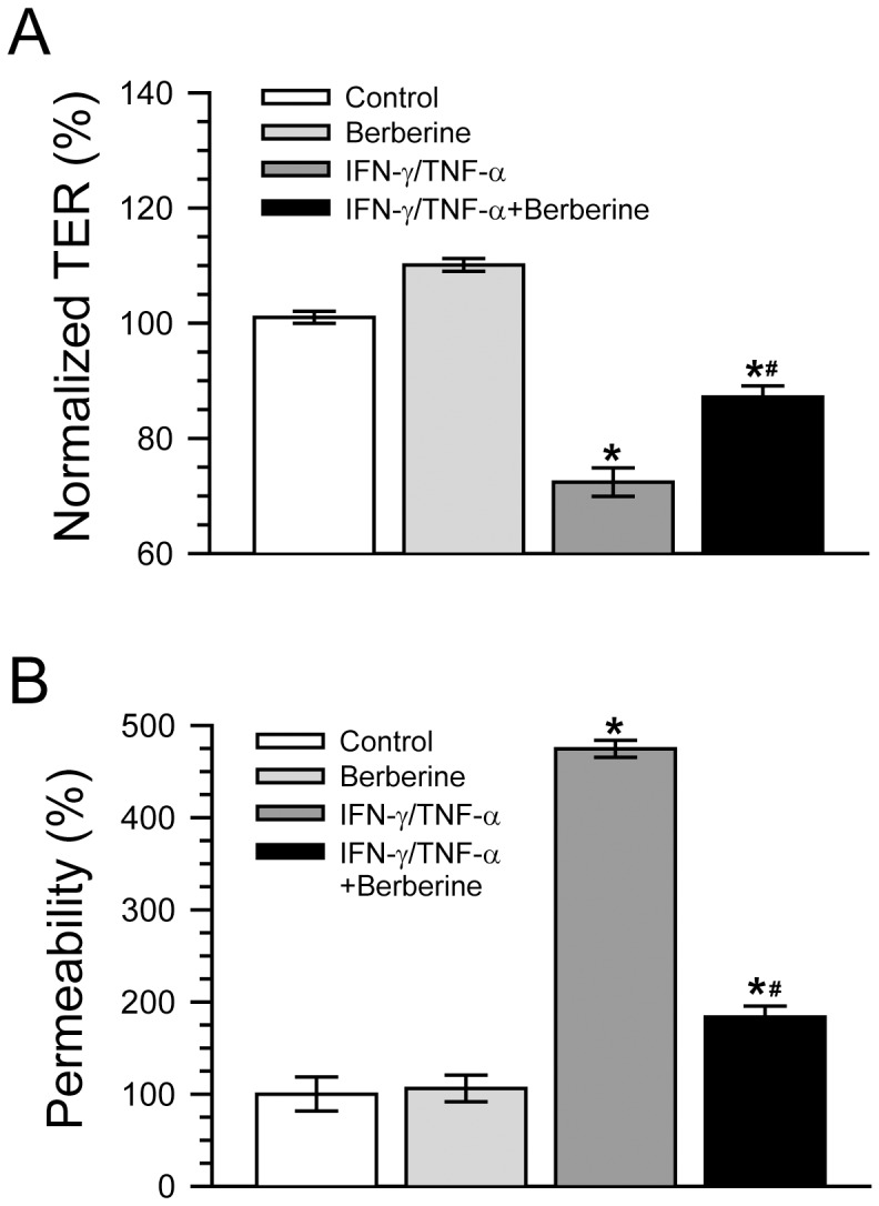
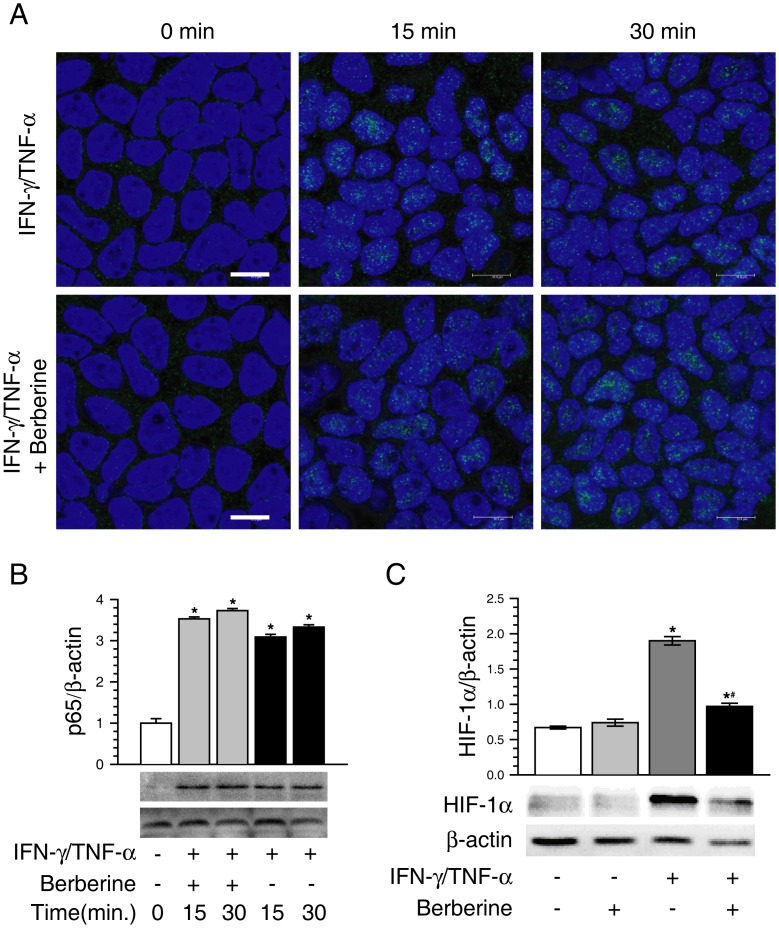
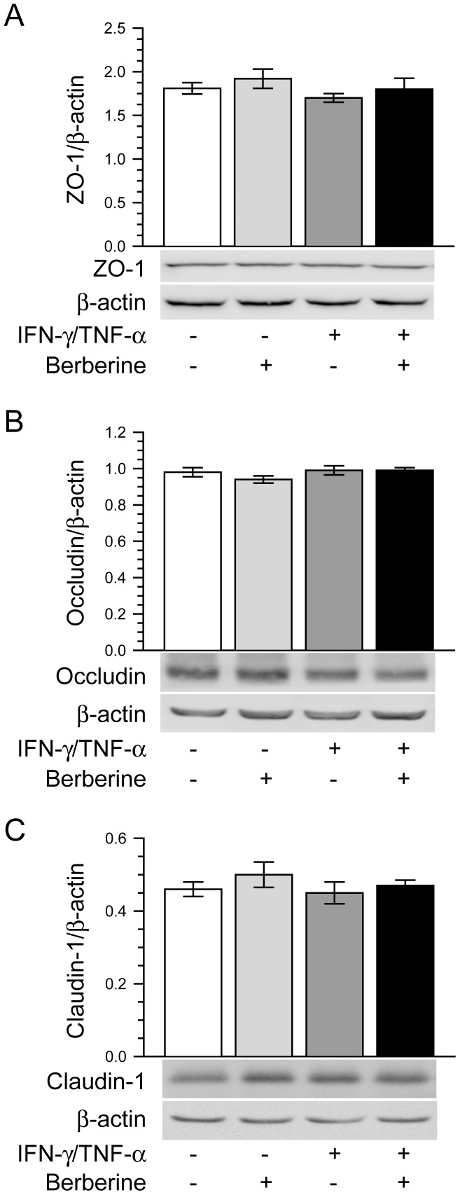
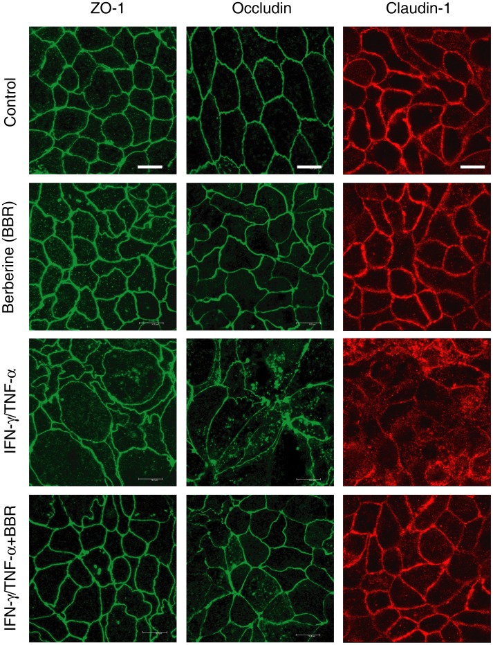
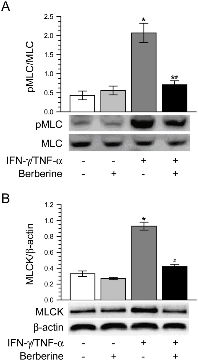
Intestinal barrier dysfunction occurs in many intestinal diseases, in which proinflammatory cytokines play critical roles. However, researchers are still on the way to defining the underlying mechanisms and to evaluate therapeutic strategies for restoring intestinal barrier function. Berberine, a drug that has clinically been used to treat gastroenteritis and diarrhea for thousands of years, has been shown to protect barrier function in both endothelial and epithelial cells, but the mechanisms are completely unknown. In this study, we investigate the protective actions of berberine on barrier function and the underlying mechanisms in Caco-2 monolayers challenged with IFN-γ and TNF-α. Caco-2 monolayers were treated without or with simultaneous IFN-γ and TNF-α in the absence or presence of berberine. Both transepithelial electrical resistance (TER) and paracellular permeability were measured to evaluate barrier function. The expression and distribution of tight junction proteins ZO-1, occluding, and claudin-1 were respectively analyzed by immunoblot or immunofluorescence. The expressions of phosphorylated myosin light chain (pMLC), MLC kinase (MLCK) and hypoxia-inducible factor-1α (HIF-1α) were determined by immunoblot. The translocation of NF-κB p65 to nuclei was analyzed by immunofluorescence and immunoblot, respectively. The results showed that berberine significantly attenuated TER decrease and paracellular permeability increase in Caco-2 monolayers treated with IFN-γ and TNF-α. Berberine also dramatically alleviated IFN-γ and TNF-α-induced morphological alteration of tight junction proteins ZO-1, occluding, and claudin-1. The increase of both MLC phosphorylation and MLCK protein expression induced by IFN-γ and TNF-α was significantly inhibited by berberine treatment. Additionally, berberine suppressed the activation of HIF-1α, but not NF-κB. Taken together, it is suggested that berberine attenuates IFN-γ and TNF-α-induced intestinal epithelial barrier dysfunction by inhibiting the signaling pathway of MLCK-dependent MLC phosphorylation mediated by HIF-1α.
肠屏障功能障碍发生在许多肠道疾病中,其中促炎细胞因子起着关键作用。然而,研究人员仍在努力确定潜在的机制,并评估恢复肠屏障功能的治疗策略。小檗碱是一种临床上用于治疗胃肠炎和腹泻已有数千年历史的药物,已被证明可保护内皮细胞和上皮细胞的屏障功能,但具体机制尚不清楚。在这项研究中,我们研究了小檗碱在 IFN-γ 和 TNF-α 攻击下对 Caco-2 单层细胞屏障功能的保护作用及其潜在机制。Caco-2 单层细胞在无或同时存在 IFN-γ 和 TNF-α的情况下进行处理,而无或同时存在小檗碱。通过跨上皮电阻(TER)和旁细胞通透性测量来评估屏障功能。通过免疫印迹或免疫荧光分别分析紧密连接蛋白 ZO-1、occluding 和 claudin-1 的表达和分布。通过免疫印迹确定磷酸化肌球蛋白轻链(pMLC)、肌球蛋白轻链激酶(MLCK)和低氧诱导因子-1α(HIF-1α)的表达。通过免疫荧光和免疫印迹分别分析 NF-κB p65 向核内的易位。结果表明,小檗碱可显著减轻 IFN-γ 和 TNF-α 处理的 Caco-2 单层细胞中 TER 降低和旁细胞通透性增加。小檗碱还显著缓解了 IFN-γ 和 TNF-α 诱导的紧密连接蛋白 ZO-1、occluding 和 claudin-1 的形态改变。IFN-γ 和 TNF-α 诱导的 MLC 磷酸化和 MLCK 蛋白表达增加均被小檗碱处理显著抑制。此外,小檗碱抑制了 HIF-1α的激活,但不抑制 NF-κB。综上所述,小檗碱通过抑制 HIF-1α 依赖性 MLCK 介导的 MLC 磷酸化信号通路,减轻 IFN-γ 和 TNF-α 诱导的肠道上皮屏障功能障碍。