Huaxi MR Research Center, Department of Radiology, West China Hospital of Sichuan University, Chengdu, PR China.
PLoS One. 2013 May 10;8(5):e63151. doi: 10.1371/journal.pone.0063151. Print 2013.
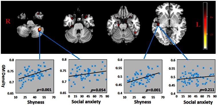
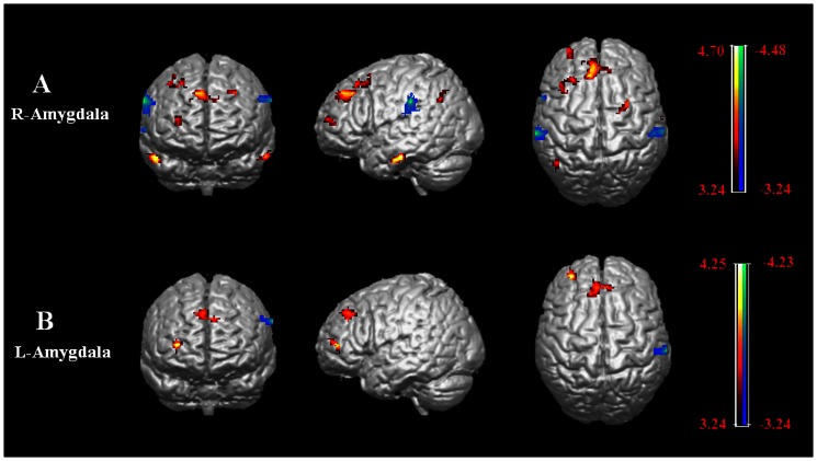
Shyness and social anxiety are correlated to some extent and both are associated with hyper-responsivity to social stimuli in the frontal cortex and limbic system. However to date no studies have investigated whether common structural and functional connectivity differences in the brain may contribute to these traits. We addressed this issue in a cohort of 61 healthy adult subjects. Subjects were first assessed for their levels of shyness (Cheek and Buss Shyness scale) and social anxiety (Liebowitz Social Anxiety scale) and trait anxiety. They were then given MRI scans and voxel-based morphometry and seed-based, resting-state functional connectivity analysis investigated correlations with shyness and anxiety scores. Shyness scores were positively correlated with gray matter density in the cerebellum, bilateral superior temporal gyri and parahippocampal gyri and right insula. Functional connectivity correlations with shyness were found between the superior temporal gyrus, parahippocampal gyrus and the frontal gyri, between the insula and precentral gyrus and inferior parietal lobule, and between the cerebellum and precuneus. Additional correlations were found for amygdala connectivity with the medial frontal gyrus, superior frontal gyrus and inferior parietal lobule, despite the absence of any structural correlation. By contrast no structural or functional connectivity measures correlated with social or trait anxiety. Our findings show that shyness is specifically associated with structural and functional connectivity changes in cortical and limbic regions involved with processing social stimuli. These associations are not found with social or trait anxiety in healthy subjects despite some behavioral correlations with shyness.
害羞和社交焦虑在某种程度上是相关的,两者都与额叶和边缘系统对社交刺激的超反应有关。然而,迄今为止,还没有研究调查大脑中是否存在共同的结构和功能连接差异可能导致这些特征。我们在一组 61 名健康成年受试者中研究了这个问题。首先,受试者根据他们的害羞程度( cheek 和 bus 害羞量表)和社交焦虑( liebowitz 社交焦虑量表)和特质焦虑进行评估。然后,他们接受了 MRI 扫描和体素形态学以及基于种子的静息状态功能连接分析,以研究与害羞和焦虑评分的相关性。害羞评分与小脑、双侧颞上回和海马旁回以及右侧脑岛的灰质密度呈正相关。与害羞相关的功能连接发现于颞上回、海马旁回和额回之间,脑岛和中央前回和下顶叶之间,以及小脑和楔前叶之间。尽管没有结构相关性,但还发现了杏仁核与额内侧回、额上回和下顶叶的连接。相比之下,没有结构或功能连接测量与社交或特质焦虑相关。我们的研究结果表明,害羞与处理社交刺激的皮质和边缘区域的结构和功能连接变化有关。这些关联在健康受试者中并未发现与社交或特质焦虑有关,尽管与害羞有一些行为相关性。